Diffuse rash spreads from infant’s scalp to extremities
A 3-month-old boy presents for evaluation of a diffuse asymptomatic rash that began on his scalp and skin creases 6 weeks ago and has spread over his trunk and extremities. This week he has begun to scratch at his neck and abdomen.
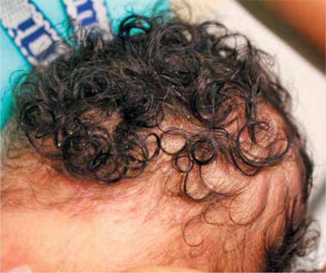
Figure 1

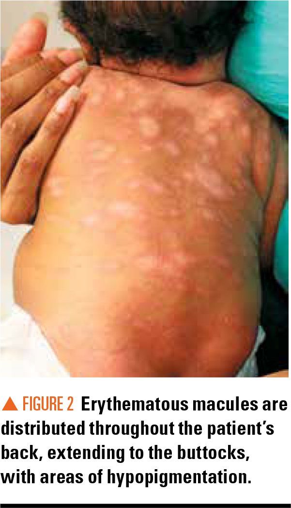
Figure 2

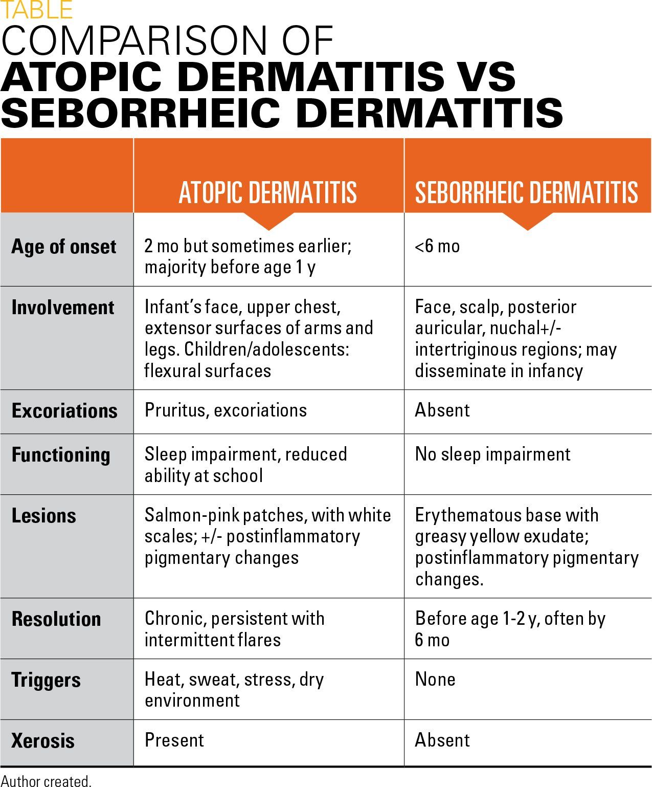
Table

The case
A 3-month-old boy presents for evaluation of a diffuse asymptomatic rash that began on his scalp (Figure 1) and skin creases 6 weeks ago and has spread over his trunk and extremities (Figure 2). This week he has begun to scratch at his neck and abdomen.
Diagnosis: Seborrheic dermatitis
Discussion
The pattern, morphology, and symptoms associated with rashes in young infants help to establish the diagnosis and treatment regimen. Atopic dermatitis (AD) and seborrheic dermatitis (SD) present commonly in early infancy and clinical features can overlap, leading to a delay in diagnosis and effective management (Table).
Seborrheic dermatitis has a prevalence of 4% in infants aged younger than 5 years.1 Within the first 3 months, SD often manifests as “cradle cap” with erythematous scaly plaques beneath a thick yellow greasy exudate. Other common sites have been coined the “seborrheic areas,” notably the face, neck, and groin. Such areas have a high density of sebaceous glands. Infant SD is not associated with significant morbidity as symptoms are often mild and self-limiting, resolving by age 12 months.2
Almost one-quarter of pediatric patients referred to pediatric dermatologists have AD.3 A majority (90%) of patients are aged younger than 5 years at presentation. Many develop symptoms within the first 3 months of life; and onset may overlap with SD. Unlike SD, AD is a chronic condition with intermittent flares that has a significant impact on a developing child’s quality of life.
Atopic dermatitis manifests as an erythematous eruption with intense pruritus. The lesions in young children tend to be moist and crusted, becoming less exudative with increasing age. This picture can be easily confused with SD, and AD may develop concurrently. Both AD and SD can be associated with hypopigmentation and/or hyperpigmentation, but lichenification, excoriations, and sparing of protected areas (eg, intertriginous and diaper areas) are typical of AD in infancy.3
The integrity of the epidermal barrier is compromised in AD, increasing risk of infection. Be aware that the motor coordination facilitating scratch does not mature until age 2 to 3 months.3 Thus, for infants aged younger than 2 to 3 months, pruritus may be more difficult to identify. Infants will manifest with irritability often writhing against clothing and stationary objects. Additionally, infants aged 2 to 3 months may also involve the forehead, cheeks, and scalp, such as SD adding to misdiagnosis, but will later spread to cover the trunk and extremities.
At this age, sparing of the intertriginous regions of the diaper may be used with caution as a distinguishing factor. The converse may also hold, that involvement of the diaper region may raise concern to consider alternative diagnoses (ie, infection or concomitant SD).
Management
Seborrheic dermatitis is a benign and self-limiting condition. The fundamental basis of treatment is emollients in order to loosen scales so they may be easily removed. In children, scalp involvement is most common and is treated with shampoos-typically twice per week-preferentially containing selenium sulfide or zinc pyrithione.4 After bathing, a fine comb may be used to remove any scales. For SD involving other areas such as the face and body, topical antifungal agents are considered first-line treatment due to the association with Malassezia yeast.
Options include ciclopirox and ketoconazole creams applied twice daily for up to 4 weeks, or the antiseborrheic shampoos containing these agents may be gently lathered on the face, ears, and neck. Alternatively, topical corticosteroids (1%-2.5% hydrocortisone) applied sparingly, not more than twice daily, and tapered as soon as possible, may clear the rash. Topical calcineurin inhibitors also may be helpful but are not approved for children aged younger than 2 years despite data demonstrating safe use in AD in children aged as young as 3 months.
Although many children with AD go into remission by elementary school, AD can be chronic with intermittent exacerbations if not controlled. Key principals in the management of AD include avoiding known triggers and treatment aimed at reducing pruritus and inflammation and maintaining barrier function. Skin hydration is paramount as the disrupted barrier leads to transepidermal water loss and xerosis.5 Daily soaking in lukewarm water, without use of any detergents or skin products, is recommended with immediate occlusive seal thereafter. This is referred to as the “soak-and-seal” method and involves application of a thick emollient after light drying to create a protective barrier.
For “hot” areas, topical corticosteroids are most effective. Hot areas are active areas during times of exacerbation. The goal is to use the lowest potency for the shortest interval in order to minimize potential adverse effects. If patients require long-term treatment, topical calcineurin inhibitors such as tacrolimus (Protopic) and the topical phosphodiesterase 4 inhibitor crisaborole may be used,5 particularly in sensitive areas such as the face and intertriginous areas. These are steroid-sparing agents that lack the adverse effects seen by long-term corticosteroid use.
The most distressing symptoms for children are the pruritus and scratching that only perpetuates these symptoms. Since the inflammatory process is primarily T-cell driven, antihistamines have not shown efficacy in controlling such symptoms. However, as skin hydration and inflammation improve with emollients and corticosteroids, the pruritus will usually decrease.
Patient outcome
The patient’s SD was treated with antiseborrheic shampoo for the scalp and a low-potency topical steroid for his face and trunk. The SD cleared but recurrent patches of AD were treated with occlusive clothing, emollients, and low-potency topical steroids.
References:
1. Schachner L, Ling NS, Press S. A statistical analysis of a pediatric dermatology clinic. Pediatr Dermatol. 1983;1(2):157-164.
2. Alexopoulos A, Kakourou T, Orfanou I, Xaidara A, Chrousos G. Retrospective analysis of the relationship between infantile seborrheic dermatitis and atopic dermatitis. Pediatr Dermatol. 2014;31(2):125-130.
3. Lapidus C, Honig PJ. Atopic dermatitis. Pediatr Rev. 1994;15(8):327-332.
4. Clark GW, Pope SM, Jaboori KA. Diagnosis and treatment of seborrheic dermatitis. Am Fam Physician. 2015;91(3):185-190.
5. Lyons JJ, Milner JD, Stone KD. Atopic dermatitis in children: clinical features, pathophysiology, and treatment. Immunol Allergy Clin North Am. 2015;35(1):161-183.

Recognize & Refer: Hemangiomas in pediatrics
July 17th 2019Contemporary Pediatrics sits down exclusively with Sheila Fallon Friedlander, MD, a professor dermatology and pediatrics, to discuss the one key condition for which she believes community pediatricians should be especially aware-hemangiomas.
Itchy skin associated with sleep problems in infants
September 27th 2024A recent study presented at the American Academy of Pediatrics 2024 National Conference & Exhibition, sheds light on the connection between skin conditions and sleep disturbances in infants and toddlers, highlighting itchy skin as a significant factor, even in the absence of atopic