Staphylococcal Scalded Skin Syndrome in a 5-Year-Old Boy
A 5-year-old boy was brought to the emergency department by his parents because of a rash that covered his entire body. The rash had started 2 days earlier, initially on the boy’s face, abdomen, and legs and had spread to his back, buttocks, and hands. There was a 1-day history of tactile fever when the child was sent home from school. He had no sick contacts and his immunizations were up-to-date. He had no significant medical history.

A 5-year-old boy was brought to the emergency department by his parents because of a rash that covered his entire body. The rash had started 2 days earlier, initially on the boy’s face, abdomen, and legs and had spread to his back, buttocks, and hands. There was a 1-day history of tactile fever when the child was sent home from school. He had no sick contacts and his immunizations were up-to-date. He had no significant medical history.
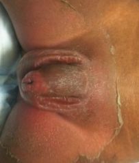
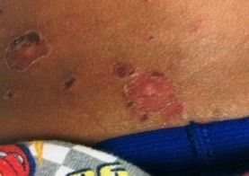
The patient appeared to be nontoxic; he was afebrile and his vital signs were stable. His skin had patches of erythema and exfoliation that were slightly tender to palpation and felt warm to the touch. The patches were distributed on his face (Figure 1, right), abdomen, back, legs, groin (Figure 2, below), buttocks, and hands.

This history and rash are classic for staphylococcal scalded skin syndrome (SSSS)-also known as staphylococcal epidermal necrolysis. This condition starts as a superficial skin blister caused by an exfoliative toxin produced by about 5% of strains of Staphylococcus aureus. The rash usually includes erythroderma of the face and of the diaper area and other intertriginous areas, and it may involve extensive areas of desquamation. SSSS spares mucous membranes, which distinguishes it from toxic epidermal necrolysis (TEN). In TEN, mucous membranes are nearly always affected (ie, the mouth, conjunctiva, trachea, esophagus, anus, vagina). It is important to differentiate SSSS from TEN. SSSS is usually benign; TEN is associated with much higher morbidity and mortality.
SSSS may be confused with bullous impetigo (Figure 3, below), another blistering skin disease caused by staphylococcal exfoliative toxins. Both conditions often manifest with a positive Nikolsky sign. In bullous impetigo, however, the toxins are restricted to the area of infection, and bacteria can actually be cultured from the blister. In SSSS, however, the toxins are spread hematogenously from a localized source, causing potential damage at distant sites. The severity of SSSS varies from a few bullae around the site of infection to severe exfoliation that affects the entire body.

SSSS is most common is young children under 6 years of age because their immune systems are immature. SSSS is rare in adults, but it has been reported in the chronically ill or immunocompromised and in those with renal failure. Complications usually result from sepsis, superinfection, and dehydration from denuded skin in severe cases; monitoring of fluid and electrolyte status is therefore important.
For children with SSSS, the prognosis is usually excellent. Immune-competent children have complete healing within 10 days without significant scarring. In adults, SSSS typically causes significant morbidity and carries high rates of mortality since most of those affected are immunocompromised with comorbidities.
Laboratory tests are not useful for diagnosis and cultures will be negative. Skin biopsies show a separation of the superficial layer of epidermis, a feature that differentiates SSSS from TEN, in which there is an epidermal-dermal separation.
Appropriate therapy depends on correct diagnosis. Antibiotics should cover for MRSA; options include cephalexin, nafcillin, or vancomycin or, for the penicillin-allergic patient, clindamycin or co-trimoxazole. Patients with severe disease should be admitted to the burn unit or ICU for parenteral antibiotic therapy, wound care, pain control, antipyretics, and close monitoring of fluid and electrolyte status. Those with mild disease can be discharged with close follow-up. As always with skin infections, encourage good hygiene and careful sanitation of common household surfaces. Discourage sharing of toiletries.
Because our patient appeared well and was able to tolerate oral medications, he was given cephalexin and co-trimoxazole and discharged home with close follow-up with his pediatrician in 1 to 2 days. His parents were encouraged to provide good oral hydration, and his family was vigilant in disinfecting the home. The patient did well without complications or recurrence, as is typical of this rare skin disease.
Suggested Reading
• Hayward A, Knott F, Petersen I, et al. Increasing hospitalizations and general practice prescriptions for community-onset staphylococcal disease, England. Emerg Infect Dis. 2008;14:720-726.
• King R. Staphylococcal Scalded Skin Syndrome in Emergency Medicine, Emedicine.com. 2010. Available at: http://emedicine.medscape.com/article/788199-overview. Accessed June 4, 2102.
• Ladhani S, Evans R. Staphylococcal scalded skin syndrome. Arch Dis Child. 1998;28:85-88.
• Patel GK, Finlay AY. Staphylococcal scalded skin syndrome: diagnosis and management. Am J Clin Dermatol. 2003;4:165-175.
Recognize & Refer: Hemangiomas in pediatrics
July 17th 2019Contemporary Pediatrics sits down exclusively with Sheila Fallon Friedlander, MD, a professor dermatology and pediatrics, to discuss the one key condition for which she believes community pediatricians should be especially aware-hemangiomas.