A 14-month old boy with coughing, fever, and failure to thrive puzzles doctors.
It's been a busy day in the emergency department (ED). You pick up the next chart and prepare to see a 14-month-old Arab American boy with a 10-day history of rhinorrhea, cough, fever, and tugging of his ear. The boy's mother explains that his pediatrician evaluated her son about 1 week ago and that the pediatrician prescribed amoxicillin for otitis media.
It's been a busy day in the emergency department (ED). You pick up the next chart and prepare to see a 14-month-old Arab American boy with a 10-day history of rhinorrhea, cough, fever, and tugging of his ear. The boy's mother explains that his pediatrician evaluated her son about 1 week ago and that the pediatrician prescribed amoxicillin for otitis media.
When his fever rose that night to 41.1°C, despite the antibiotics, she took her son the next day to a community hospital ED, where a blood culture and complete blood count (CBC) with differential were drawn. Fortunately, the mother has brought a copy of the records from that ED visit.
The CBC showed a white blood count (WBC) of 25,000 cells/mm3, with a differential of 57% neutrophils, 31% lymphocytes, 11% monocytes, and no bands. Hemoglobin (Hb) was 8 g/dL; platelet count was 553,000 cells/mm3; and the urinalysis was normal. Nasal washes for respiratory syncytial virus (RSV) and influenza were negative.
The boy was given 1 dose of intramuscular ceftriaxone and then sent home but was called back to the pediatrician’s office because of growth in the blood culture obtained in the ED. The mother tells you that the pediatrician repeated the CBC and blood culture and said that the WBC had come down. She says the pediatrician thought the blood culture result was just a contaminant.
You note that the ED paperwork shows that the blood-culture isolate was identified as Staphylococcus warneri, a coagulase-negative organism that commonly colonizes the skin, so this seems consistent. The mother tells you that the repeat blood culture drawn in the pediatrician’s office was negative. The patient, however, continued to have intermittent fevers that were treated with acetaminophen and ibuprofen for approximately 3 weeks. Frustrated and concerned, the mother decided to bring her son to the children’s hospital ED for further evaluation.
History
You ask about past medical history and elicit that the boy was born full term, weighing 3.422 kg (25th-50th percentile) via spontaneous vaginal delivery to a healthy 21-year-old woman who had no pregnancy complications and reports unremarkable prenatal laboratory results. The mother tells you that she tested negative for human immunodeficiency virus (HIV), hepatitis B, and syphilis during the first trimester of pregnancy, and you verify this by reviewing the mother’s prenatal records.
The child was breastfed for 2 weeks after delivery then was switched to cow’s milk formula because the mother felt she did not have an adequate milk supply. His length and head circumference at birth were measured in the 25th percentile, and his mother has been concerned for some time about his eating and his growth; however, he has had no developmental delay or loss of milestones. The child was born in the United States and has received all age-appropriate immunizations. He lives with his parents, both of whom were born outside the United States. The child has no siblings. He has had no previous hospitalizations.
Physical findings
The child is alert and crying but consolable. His temperature is 39.1°C (rectal); weight is 8.8 kg (<3rd percentile); height is 73 cm (<5th percentile); head circumference is 45.1 cm (5th percentile); heart rate is 120 beats per minute; respiratory rate is 26 breaths per minute; and blood pressure is 100/54 mm Hg.
The otoscopic exam reveals an erythematous and bulging right tympanic membrane. His oropharynx is noninjected, and there is no thrush. There are a few scattered crackles heard bilaterally. Heart exam reveals a regular rate and rhythm without murmurs, abdomen is soft without hepatosplenomegaly, and capillary refill time is 2 seconds. No rashes or lesions are noted on skin examination.
The musculoskeletal exam reveals full range of motion, adequate strength, and no erythema or effusions. Neurologically, his deep tendon reflexes are 2/4 bilaterally, and cranial nerves II to XII are symmetric without any focal deficits. The positive blood culture from the outside hospital and the persistent fevers despite antibiotic therapy prompt you to do further workup. You admit him to the children’s hospital for further observation. For your initial differential diagnosis, you consider bacteremia, viral infection, and drug-resistant otitis media.
A plan of action
You draw blood for culture and CBC, obtain urine for culture and nasal wash for influenza and RSV, and order a chest x-ray. The CBC shows a WBC of 23,900 cells/mm3, with a differential of 9% bands, 36% neutrophils, and 47% lymphocytes; Hb of 8.8 g/dL; and platelet count of 232,000 cells/mm3. Nasal washes for influenza A and B are negative, but the RSV nasal wash is positive. There is no infiltrate or pleural effusion. The chest x-ray reading indicates small airway disease. Because of the persistent acute otitis despite oral antibiotics and concern for bacteremia, you continue the ceftriaxone.
Blood and urine cultures are sterile after 2 days. The CBC shows a decline in the WBC count to 17,900 cells/mm3. The Hb remains low at 8.1 g/dL. The reticulocyte count is 5.6%. The child remains afebrile during his hospital stay. Iron studies return and are not consistent with a diagnosis of iron-deficiency anemia. The mean corpuscular volume is 81.8 fL (normocytic), and the red blood cells are normochromic on smear.
You note that his mother is thin in build. The boy’s diet consists of milk and cereal with no meat or other sustainable form of protein. As you look through his records, you note that even though at birth his weight was between the 25th and 50th percentile, he has steadily been crossing percentiles to less than the 3rd percentile during this admission. His height was following the 10th percentile but has now fallen in the last year to the 3rd percentile.
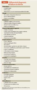
You are concerned that the boy’s failure to thrive (FTT) is not fully explained by poor nutrition and obtain further lab studies. Electrolyte levels along with blood urea nitrogen, creatinine, calcium, magnesium, phosphorus, and bilirubin levels are normal. Alanine and aspartate aminotransferases are elevated at 260 units/L and 450 units/L, respectively. Alkaline phosphatase, lactate dehydrogenase (LDH), uric acid, and soluble transferrin receptor levels are normal. You think of the differential for FTT again (Table 1)1 and consider the transaminitis, history of persistent fevers, otitis media resistant to oral therapy, and the normocytic anemia. The FTT does not appear to be purely nutritional. The child is afebrile at discharge and given a follow-up appointment to continue the FTT evaluation.

Back into the hospital
Before his appointment, he returns to the ED with fever, diarrhea, and decreased oral intake. You are informed that although the child’s fever had resolved during his previous hospital admission, it returned 1 week after he was discharged. Today, the CBC with differential shows an elevated WBC of 18,600 cells/mm3 with 3% bands, 44% neutrophils, and 44% lymphocytes. A repeat blood culture is also sent. Alanine and aspartate aminotransferases have risen even further to 524 units/L and 526 units/L, respectively.
On physical exam he does not appear toxic. There are no abnormal findings on exam, and the otitis media has resolved. The child is febrile with a temperature of 39.7°C. You decide to readmit the patient.
During this admission, you delve further into the social history and past medical history of this child. You ask about past or family history of metabolic abnormalities, celiac disease, autoimmune diseases, consanguinity, or history of transfusions for either the child or mother during pregnancy. The history for each is negative. Alpha-1-antitrypsin, alpha-1-phenotype, hepatitis B surface antigen and antibody, and hepatitis C antibody screen are all negative. An abdominal ultrasound shows mild hepatomegaly. In the past month, the patient has gained only 100 g, from 8.8 kg to 8.9 kg.
On exam, the liver is palpable at 5 cm below the right costal margin. The hepatitis screen is negative. In order to determine whether there is an underlying immune deficiency to explain the FTT, repeated febrile illnesses, and infections, you send for tests for immunoglobulins (IgG, IgA, IgM, IgE), lymphocyte subsets, and antibody titers to pneumococcus, diphtheria, and tetanus. You review the case with the hematologist, who recommends testing for glucose-6 phosphate deficiency (G6PD) as a cause of the anemia. He is again given intravenous antibiotics, the fever and diarrhea resolve, and the child is discharged home with a follow-up appointment in clinic.
The follow-up
The child returns for follow-up at your clinic. His laboratory results reveal G6PD deficiency that appears to confirm the etiology of his anemia. The IgG is elevated to 2,810 mg/dL (normal range, 345-1,213 mg/dL), but the IgM and IgA are within normal parameters. You attribute the elevated IgG to recent infection. You are concerned because of the child’s insufficient weight gain since his hospital discharge.
You once aga
in ask about the home situation and state of health of other members of the family. The mother reports that her primary care doctor is evaluating her for lymphadenopathy. Considering this new information, you realize that in this child’s previous evaluation for FTT, an HIV screen was never performed. You recall that his mother had a negative HIV enzyme-linked immunosorbent assay (ELISA) during the first trimester of her pregnancy, but you decide to order the HIV DNA polymerase chain reaction (PCR) test as well as an HIV ELISA on this child.
The HIV ELISA and subsequent Western blot for HIV come back positive, which confirms perinatal exposure to HIV. The HIV DNA PCR returns 3 days later, confirming the child’s HIV infection. You refer the patient to immunology for further care and evaluation.
Failure to thrive
Failure to thrive is better described as a failure to gain weight.1 Most commonly, this is because of inadequate nutritional intake. However, other causes include inadequate calorie intake, inadequate caloric absorption, and excessive caloric expenditure, which can be because of conditions such as congenital heart disease, pulmonary disease, hyperthyroidism, metabolic disease, chronic immunodeficiency, recurrent infection, or malignancy.2 The proportion of children seen in the primary care setting with FTT has been found to be as high as 10%. Failure to thrive can be the presenting finding in children with perinatal HIV infection.3
Pediatric HIV
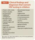
Many clinical problems, including FTT, warrant HIV testing in their workup (Table 2).4 HIV in infants became uncommon with the implementation of treatment guidelines established after the landmark 076 study of the AIDS Clinical Trial Group in 1994, whereby all women are offered HIV testing during pregnancy, all infected women receive antiretroviral drugs during pregnancy and during labor and delivery, and known infants exposed to HIV receive oral zidovudine for the first 6 weeks after birth.5,6 With use of combination antiretroviral drugs during pregnancy now routine, the incidence of pediatric HIV infection has plummeted.

The primary mode of infant HIV acquisition is mother-to-child transmission (MTCT) via transplacental, intrapartum, or breastfeeding routes of exposure. Perinatally acquired HIV-1 infection shows 2 distinct patterns if untreated. During the first few weeks of life, 10% to 15% of infected infants have a profound immune deficiency leading to opportunistic infections, usually pneumocystis pneumonia.3 The other pattern is characterized by more gradually progressive immune deficiency over a period of several years and manifesting as lymphadenopathy, poor growth, and repeated bacterial infections.
Studies on timing of MTCT of HIV suggest that most transmissions in nonbreastfeeding populations occur at the time of delivery.7 The risk of transmission is particularly high for women with a primary HIV-1 infection during the last trimester of pregnancy. These women are more likely to have higher viral loads and less likely to have their HIV infection identified through usual HIV antibody testing.
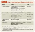
The method of diagnosis for HIV varies by age group (Table 3).4 In children aged 18 months and older and in all adolescents and adults, HIV antibody is the confirmatory test. Most commonly, a blood sample is used, and testing is done via ELISA testing with confirmation using a Western blot. Plasma HIV RNA concentration (by PCR), or viral load, is used to determine the need for treatment and for monitoring response to treatment.

In infants aged through 18 months and for anyone who cannot make antibody (eg, a patient with common variable immune deficiency), HIV DNA (or RNA) by PCR is used to diagnose HIV.4 Antibody testing in infants younger than 18 months is not useful, because a positive test indicates the presence of passively acquired maternal antibody in the infant’s serum and does not distinguish HIV-infected from HIV-exposed, uninfected infants.
HIV testing
Current CDC guidelines recommend early HIV testing for all pregnant women, with repeat testing in the third trimester in women who meet at least 1 of the following criteria: 1) Receive health care in jurisdictions with elevated incidence of HIV or AIDS among women aged 15 to 45 years; 2) Receive health care in facilities in which prenatal screening identifies at least 1 HIV-infected pregnant woman per 1,000 women screened; 3) Known to be at high risk for acquiring HIV (eg, injection-drug users and their sex partners; exchange sex for money or drugs; are sex partners of HIV-infected persons; have had a new or more than 1 sex partner during this pregnancy); and 4) Have signs or symptoms consistent with acute HIV infection.8
This patient’s mother was not identified through the usual perinatal screening (a missed opportunity to prevent transmission), and the child’s diagnosis was delayed because of several factors. Such presentations of pediatric HIV infection now are uncommon in the United States because of widespread HIV testing of pregnant women and implementation of effective strategies to prevent MTCT for women known to have HIV infection. She was tested for HIV during pregnancy and was negative in the first trimester but did not receive repeat testing later in the pregnancy. She did not meet the high-risk criteria for repeat testing. She may have been in the window period when initially tested for HIV or may have acquired HIV infection during her second or third trimester. This woman was not considered to be a high-risk patient for contracting HIV because she reported only 1 lifetime partner and her ethnic background (Arab American) is not considered to be in the high endemic group of her state. She was not living in an area that was considered high enough prevalence for repeat testing during third trimester or at delivery.
Several states, including Michigan (where this infant was born) are reporting cases of infants and toddlers with HIV who were missed at birth because of first-trimester negative HIV ELISA without repeat testing in the third trimester or at delivery.9 Because of this case and others, the policy in Michigan has been changed to include a recommendation for third-trimester testing or rapid testing during delivery.10
Initial laboratory testing in an HIV-infected patient should include CD4 percentage and absolute cell counts, plasma HIV RNA concentration (viral load), and the HIV genotype to assess for baseline resistance mutations.4 In children aged younger than 5 years, the CD4 percentage is the preferred test for monitoring immune status because the absolute CD4 cell count (number of CD4 cells/mm3) in this age group varies with age-related changes in the absolute lymphocyte count.
Our patient
The patient was seen in the pediatric HIV clinic. The CD4+ T-cell count was 2,432 (38%), which was normal for age (2,307-2,864), and the HIV viral load was found to be 134,000 copies/mL. The genotyping of the virus did not reveal any resistance to antiretroviral drugs, and he was started on antiretroviral therapy because the viral load was above 100,000 copies/mL. The drug regimen consisted of zidovudine, lamivudine, and nevirapine. Both parents were confirmed to be HIV positive. On the return visit 2 months later, the patient had a CD4+ T-cell count of 2,500 and an HIV viral load of 115 copies/mL. Approximately 7 months into treatment, the patient’s viral load was less than 48 copies/mL (undetectable).
References
- McLean HS, Price DT. Failure to thrive. In: Kleigman RM, Stanton BF, St Geme JW III, Schor NF, Behrman RE, eds. Nelson Textbook of Pediatrics. 19th ed. Philadelphia, PA: Elsevier Saunders; 2011:147-149.
- Stephens MB, Gentry BC, Michener MD, Kendall SK, Gauer R. Clinical inquiries. What is the clinical workup for failure to thrive? J Fam Pract. 2008;57(4):264-266.
- Seeborg FO, Paul ME, Abramson SL, et al. A 5-week-old HIV-1-exposed girl with failure to thrive and diffuse nodular pulmonary infiltrates. J Allergy Clin Immunol. 2004;113(4):627-634.
- Simpkins EP, Siberry GK, Hutton, N. Thinking about HIV infection. Pediatr Rev. 2009;30(9):337-348.
- Frenkel LM, Cowles MK, Shapiro DE, et al. Analysis of the maternal components of the AIDS clinical trial group 076 zidovudine regimen in the prevention of mother-to-infant transmission of human immunodeficiency virus type 1. J Infect Dis. 1997;175(4):971-974.
- Connor EM, Sperling RS, Gelber R, et al. Reduction of maternal-infant transmission of human immunodeficiency virus type 1 with zidovudine treatment. Pediatric AIDS Clinical Trials Group Protocol 076 Study Group. N Engl J Med. 1994;331(18):1173-1180.
- American Academy of Pediatrics. Human immunodeficiency virus infection. In: Pickering LK, Baker CJ, Kimberlin DW, Long SS, eds. Red Book: 2009 Report of the Committee on Infectious Diseases. 28th ed. Elk Grove Village, IL: American Academy of Pediatrics; 2009:380-400.
- Branson BM, Handsfield HH, Lampe MA, et al; Centers for Disease Control and Prevention (CDC). Revised recommendations for HIV testing of adults, adolescents, and pregnant women in health-care settings. MMWR Recomm Rep. 2006;55(RR-14):1-17.
- Faghih S, Secord E. Increased adolescent HIV infection during pregnancy leads to increase in perinatal transmission at urban referral center. J Int Assoc Physicians AIDS Care (Chic). 2012;11(5):293-295.
- Michigan Department of Community Health. Guidelines for testing and reporting: perinatal human immunodeficiency virus (HIV), hepatitis B and syphilis. Revised August 31, 2010. www.michigan.gov/documents/mdch/HIV_Hep_B_Syphilis_Guidelines_forTesting_and_Reporting_083_205_331936_7.pdf. Accessed December 3, 2012.