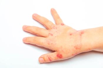
Botfly Infestation in a Teenage Boy
An infected insect bite was initially diagnosed, and a course of oral trimethoprim/sulfamethoxazole was started. Eight days later, the patient returned with worsening symptoms and a "white thing poking in and out" of one of the lesions (A). He was advised to occlude the lesion with petroleum jelly and an adhesive bandage. The next day, the patient brought in the "creature" that had emerged from the lesion. It was subsequently identified as a larva of the human botfly, Dermatobia hominis. A second larva emerged from the other lesion 1 week later.
Figure
FigureFive weeks after returning from a school trip to the Amazon in Peru, a 16-year-old boy presented with 2 persistent "bug bites" on the lateral left calf. He had sustained multiple bug bites during his trip, all of which had resolved spontaneously except for the 2 on his lower leg. These were associated with intermittent bleeding, pruritus, and sharp pain.
The 2 2-cm, erythematous, firm nodules had a central punctum and clear discharge. All other physical findings were normal.
An infected insect bite was initially diagnosed, and a course of oral trimethoprim/sulfamethoxazole was started. Eight days later, the patient returned with worsening symptoms and a "white thing poking in and out" of one of the lesions (A). He was advised to occlude the lesion with petroleum jelly and an adhesive bandage. The next day, the patient brought in the "creature" that had emerged from the lesion. It was subsequently identified as a larva of the human botfly, Dermatobia hominis. A second larva emerged from the other lesion 1 week later.
Myiasis is caused by larvae of the order Diptera, the 2-winged true fly.1 Furuncular myiasis begins as pruritic papules that evolve into boillike lesions with central punctums. D hominis is the most common causative organism in Central and South America. Mosquitoes transfer the botfly eggs to mammalian hosts via a bite. Local heat causes the eggs to hatch.2 The larvae burrow underneath the epidermis, where they feed and grow for about 5 to 10 weeks until they eventually tunnel out of the skin and drop to the soil, where they pupate (B). The effects on human hosts are mostly minimal but can include local sensations of gnawing and drilling, regional lymphadenopathy, fever, and anxiety. Complications include bacterial superinfection and tetanus.
Furuncular myiasis is often mistaken for a furuncle. 3,4 However, it is easily differentiated by the lack of response to antibiotic therapy and by the emergence of a larva through the central punctum. Ultrasonography may help determine whether a larva is present.5
In Central and South America, where the disease is endemic, pork fat is used to obstruct the pore; the fat cuts off the larva's oxygen supply, forcing it out of its burrow.6 Other occlusive materials, such as petroleum jelly and adhesive tape, are also effective.2 After the larva emerges, it can be carefully removed with forceps. Surgical removal is considered in special cases, such as those with palpebral involvement. Incorrect or partial removal of the larva can lead to secondary infection.4
Furuncular myiasis can be prevented by using mosquito nets, wearing clothing that covers the extremities, and applying insect repellent.2
References:
- Jacobson CC, Abel EA. Parasitic infestations. J Am Acad Dermatol. 2007;56: 1026-1043.
- Mathieu ME, Wilson BB. Myiasis and tungiasis. In: Mandell GL, Bennett JE, Dolin R, eds. Principles and Practices of Infectious Disease. 6th ed. Philadelphia: Churchill Livingstone; 2005:3307-3310.
- Maier H, Honigsmann H. Furuncular myiasis caused by Dermatobia hominis, the human botfly. J Am Acad Dermatol. 2004;50:S26-S30.
- Bhandari R, Janos DP, Sinnis P. Furuncular myiasis caused by Dermatobia hominis in a returning traveler. Am J Trop Med Hyg. 2007;76:598-599.
- Quintanilla-Cedillo MR, Leon-Ureña H, Contreras-Ruiz J, Arenas R. The value of Doppler ultrasound in diagnosis in 25 cases of furunculoid myiasis. Int J Dermatol 2005;44:34-37.
- Brewer TF, Wilson ME, Gonzalez E, Felsenstein D. Bacon therapy and furuncular myiasis. JAMA. 1993;270:2087-2088.
Newsletter
Access practical, evidence-based guidance to support better care for our youngest patients. Join our email list for the latest clinical updates.


