Chlamydia pneumoniae: An elusive pathogen
C pneumoniae is a frequent cause of respiratory disease in children, but pinning down a laboratory diagnosis is difficult. Here are some tips on when to suspect this organism and how to treat the infection.
Chlamydia pneumoniae: An elusive pathogen
By Margaret R. Hammerschlag, MD
C pneumoniae is a frequent cause of respiratory disease in children,but pinning down a laboratory diagnosis is difficult. Here are some tipson when to suspect this organism and how to treat the infection.
Chlamydia pneumoniae infection is a puzzle for clinicians. Definitivediagnostic tests are not readily available. Some infected children are clearlyill, while others have no discernible symptoms. And when infected childrenare symptomatic, one can't be sure that C pneumoniae is responsible; co-infectionwith other organisms is common.
I wish I and other researchers in this field had answers to these clinicalconundrums, but that is not the case. The material that follows will reviewwhat we know about the peculiar physiology of this organism, explain whymaking a positive identification is so difficult, and bring you up-to-dateon the status of diagnostic testing. Finally, an epidemiologic approachto treatment will be suggested.
An important respiratory pathogen
The genus Chlamydia is a group of obligate intracellular parasites thathave a unique developmental cycle with morphologically distinct infectiousand reproductive forms. The genus contains four recognized species: Chlamydiatrachomatis, Chlamydia psittaci, C pneumoniae, and Chlamydia pecorum.C trachomatisis a well-known sexually transmitted pathogen; C psittaci and C pecorumare of great veterinary importance and the former is the agent of psittacosisin humans.
C pneumoniae, first described in 1986, has by now been recognized asan important respiratory pathogen in humans, affecting all ages.1It has been associated with community-acquired pneumonia, bronchitis, pharyngitis,otitis media, and exacerbations of asthma--all disorders of considerableinterest to pediatricians. Recent studies have also implicated C pneumoniaeinfection as a possible contributing factor in atherosclerosis.
The physiology of C pneumoniae has a direct impact on how these infectionsare diagnosed and treated. All members of the Chlamydia genus have a gram-negativeenvelopewithout peptidoglycan, share a genus-specific lipopolysaccharide antigen,and use host adenosine triphosphate (ATP) for the synthesis of chlamydialprotein.
The chlamydial developmental cycle includes an infectious, metabolicallyinactive extracellular form called an elementary body, and a noninfectious,metabolically active intracellular form called a reticulate body. Elementarybodies, which are 200 to 400 mmin diameter, attach to the host cell by electrostaticbinding and are taken into the cell by endocytosis. Within the host cell,the elementary body remains within a membrane-lined phagosome and does notfuse with the host cell lysosome.
After elementary bodies enter the cell, they differentiate into reticulatebodies, which undergo binary fission and form intracytoplasmic inclusions.The organism is identified in tissue culture by the presence of these inclusions,which may be thought of as microcolonies.
After approximately 36 hours, the reticulate bodies differentiate intoelementary bodies and infectivity increases. During this process, cell divisioncontinues unimpeded. At about 48 hours, release of the mature chlamydialelementary bodies may occur by cytolysis, or by exocytosis or extrusion,leaving the host cell intact to begin the cycle again. This sequence ofevents, in which the host cell is left relatively intact and cell divisionunaffected, accounts for the prolonged, often asymptomatic character ofChlamydia infection. This life cycle is also very long compared to conventionalbacteria, which have a doubling time in broth media of about 30 minutes.Diagnosis requires use of tissue culture and treatment often requires long,multiple dose regimens.
Who gets infected?
C pneumoniae appears to be mainly a human pathogen. It has been isolatedfrom koalas in Australia, from horses, and recently from a frog, but whetheran animal reservoir exists is not known. The mode of transmission remainsuncertain, but infected respiratory secretions are the most probable route.C pneumoniae can remain viable on formica counter tops for 30 hours andcan survive small particle aerosolization.2 The infection oftenspreads within families and enclosed populations such as military recruits.3,4
Studies of the role of C pneumoniae in community-acquired pneumonia inchildren show considerable variation, from no evidence of C pneumoniae infectionat all to evidence in as many as 18% of cases, depending on the geographiclocation and the diagnostic method used.57 Most of thesestudies relied entirely on serology, and the sero-epidemiologic data theyyielded led researchers to believe respiratory infection due to C pneumoniaewas rare in children under 5 years of age because they generally did nothave antibody.
However, recent studies using culture have found a poor correlation betweenculture and serology, especially in children. These newer studies have foundthat infection with C pneumoniae occurs as frequently in young childrenas in adults. As part of a multicenter pneumonia treatment study in children3 to 12 years of age, Block and colleagues isolated C pneumoniae from 34of 260 children (13.1%) enrolled.5 Serologic evidence of acuteinfection was found in 48 (18.5%), but only eight of the culture-positivechildren (23%) met the serologic criteria for acute infection. In a subsequentmulticenter study, Harris and colleagues isolated C pneumoniae from 6.7%of 456 children, 6 months through 16 years of age, with community-acquiredpneumonia.7 Only five of the 31 culture-positive children (16%)met the serologic criteria for acute infection; most were seronegative.In both studies, the prevalence of culture-documented C pneumoniae infectionwas the same in the children who were less than 6 years of age as it wasin those who were older.
Coinfections with other organisms were frequent. Block and colleaguesfound that almost 20% of the children with C pneumoniae infection were alsoinfected with Mycoplasma pneumoniae.5 Clinically, these childrencould not be differentiated from those who were infected with either organismalone. One child who was culture-positive for C pneumoniae was also theonly child in the study with pneumococcal bacteremia. Coinfections withStreptococcus pneumoniae has also been reported frequently in adults withC pneumoniae infection. C pneumoniae can interfere with mucociliary clearance,which may pave the way for a secondary bacterial infection.8Cultures may remain positive from several weeks to several years after acuteinfection.9,10 Asymptomatic carriage of C pneumoniae occurs in2% to 5% of adults and children, but the role it plays in the epidemiologyof C pneumoniae infection is not known.1113 Individualswith asymptomatic infection may be a reservoir for spread of infection.
Clinical presentation not very distinctive
Most respiratory infections due to C pneumoniae are probably mild orasymptomatic. Initial reports emphasized a mild, "atypical" pneumoniaclinically resembling that associated with M pneumoniae. In subsequent studies,however, pneumonia associated with C pneumoniae has clinically been indistinguishablefrom pneumonias caused by other bacteria. Coinfection with other pathogensfurther complicates the clinical diagnosis.
Occasionally, C pneumoniae has been associated with severe illness andeven death, although the role of preexisting chronic conditions in thesecases is difficult to assess. In some, C pneumoniae is clearly implicatedas a serious pathogen even in the absence of underlying disease.
Association with other disorders common
While the role of host factors in C pneumoniae infection has not beenpinned down, the infection has been identified in children with a varietyof disorders.
Asthma. One of the most exciting findings in C pneumoniae research isthe association of the organism with reactive airway disease. Many infectiousagents, including M pneumoniae and respiratory viruses, have been implicatedas inflammatory triggers for asthma. In 1989, we saw a patient with culture-documentedC pneumoniae infection who developed significant bronchospasm.9She was diagnosed with asthmatic bronchitis and was receiving systemic andinhaled steroids. She did not improve until her chlamydial infection wastreated.
Other studies have reported an association between serologic evidenceof C pneumoniae infection and wheezing in adults, although isolation ofthe organism was not done.14 The experience with children isdifferent. We were able to isolate C pneumoniae from 13 of 118 children5 to 15 years of age (11%) who were being evaluated for new asthma or acuteexacerbations of chronic asthma.12 Eradicating the organism ledto clinical improvement and better pulmonary function test scores.
Only five of the children with confirmed infection had detectable IgGantibody to C pneumoniae. One child who was noncompliant with his antibiotictherapy was culture-positive on five occasions over a three-month period.No anti-C pneumoniae antibody was ever detected. However, specific anti-Cpneumoniae IgE was found in 85.7% of the culture-positive asthmatics comparedto 9% of children with C pneumoniae pneumonia who were not wheezing.15This suggests that bronchial reactivity seen with C pneumoniae infectionmay be IgE-mediated. A similar association was seen in the patients withcystic fibrosis (CF) and C pneumoniae infection.
C pneumoniae may be especially well suited to be an infectious triggerfor asthma. It can cause prolonged, persistent infection, which produceschronic inflammation that may trigger bronchospasm in susceptible individuals.Steroid treatment may, in fact, exacerbate bronchospasm if C pneumoniaeis present. Steroids have been shown to enhance the growth of C pneumoniaein vitro and to reactivate pulmonary infection in animals.16,17
Otitis.C pneumoniae has been isolated from middle ear fluids of childrenand adults with otitis media. We recently found the organism in the middleear fluids of eight of 101 children(8%) with acute otitis media.13The infected children ranged in age from 16 to 59 months, with a mean ageof 16 months. The organism was isolated from both ears in two children andwas the only pathogen isolated from another two. Copathogens in the remainingsix children were penicillin-resistant S pneumoniae,b-lactamase positiveHaemophilus influenzae, and Moraxella catarrhalis. Although five of thechildren with C pneumoniae infection responded to a single course of treatmentwith b-lactam antibiotics, three required multiple courses of b-lactam antibioticsover several weeks. As all thebacterial isolates were susceptible to theantibiotics used in these three children, C pneumoniae coinfection may haveplayed a role in their refractory course.
Sickle cell disease. C pneumoniae appeared to be responsible for sixof 31 episodes (19%) of acute chest syndrome in children with sickle celldisease.18 As all these children had received at least one doseof pneumococcal vaccine and most were still on penicillin prophylaxis, therewere no documented pneumococcal infections. C pneumoniae infection in thesepatients was associated with more severe hypoxia than infection with M pneumoniae.
Cystic fibrosis. We have also isolated C pneumoniae from four of 32 patients(12.5%) with CF presenting with acute pulmonary exacerbations and failedto isolate the organism from 24 clinically stable patients with CF.19One patient, a 41 year-old woman, evidently acquired the infection fromher children, who had symptoms of an upper respiratory infection one weekprior to her illness. As survival rates of children with CF lengthen, thesusceptibility of these individuals to community-acquired respiratory pathogenssuch as C pneumoniae becomes a matter of concern.
HIV infection. Children with HIV infection may also be susceptible torespiratory illness caused by C pneumoniae. The organism was isolated fromfour of 22 HIV-positive children (18%) with new onset of pneumonia, andappeared to be the sole cause of the pneumonia.20
Laboratory diagnosis difficult
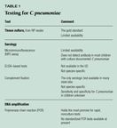
Making a positive identification of C pneumoniae is fraught with difficulty.Three approaches are possible: culture, serology, and DNA amplification(Table 1). None of the tests based on these approaches is readily availableat present. Often, clinicians will have to be content with empirical approachesto treatment.

Culture. At present, culture of swab specimens from nasopharyngeal orpleural fluid, if available, is the only reliable way to make a laboratorydiagnosis. The nasopharynx appears to be the best site.5 Cultureon HEp-2 cells with an initial inoculation and one passage should take fourto seven days. Unfortunately, culture is not widely available outside asmall number of research laboratories.
To get a culture diagnosis, obtain a nasopharyngeal specimen with a Dacron-tippedwire-shafted swab. Place the specimen in an appropriate transport medium,usually a sucrose-phosphate buffer with antibiotics and fetal calf serum,and store it immediately at 4° C for no longer than 24 hours. Viabilitydecreases if specimens are held at room temperature. If the specimen cannotbe processed within 24 hours, freeze it at 70° C until cell culturecan be performed. After 72 hours incubation, the laboratory can confirmthe diagnosis by staining with either a C pneumoniae species-specific ora Chlamydia genus-specific (anti-LPS) fluorescein-conjugated monoclonalantibody.21
Serology. Because culturing C pneumoniae is not readily available orperformed, researchers continue to pursue a serologic diagnosis--predominantlywith the microimmunofluorescence (MIF)test. Unfortunately, the MIF testis also limited to a small number of research laboratories and is not standardized.Two MIF kits (MRL and LabSystems) are available and are used by some largelaboratories. Neither has been approved by the FDA.
ELISAbased serologic tests have also been developed. They are genus-specificand are not available in the US.
The only Chlamydia serology available to most American pediatriciansis a complement fixation test. It is genus-specific and used mainly forthe diagnosis of psittacosis. Sensitivity and specificity for C pneumoniaeinfection in children is unknown; none of the C pneumoniae culture-positivechildren reported by Block and colleagues had any detectable antibody bythis test.5
Because a serologic response to primary infection takes some time todevelop, it may be missed if convalescent sera are obtained earlier thanthree weeks after the onset of illness. Use of paired sera taken duringthe acute and convalescent stages of illness affords only a retrospectivediagnosis, of little help in deciding how to treat the patient. The criteriafor use of a single serum sample have not been correlated with the resultsof culture and are based mainly on data from adults. Most children withculture-documented C pneumoniae infection do not have antibody detectableby the MIF assay. Of the culture-positive children enrolled in the multicenterpneumonia treatment study, most had no detectable antibody by the MIF testeven after three months of follow-up.5 Immunoblottesting revealedthat these children had antibody to a number of C pneumoniae proteins, butfewer than 30% of the children reacted with the antigen presented in theMIF test.22
DNA amplfication. The polymerase chain reaction (PCR) appears to be themost promising technology for developing a rapid, nonculture method fordetecting C pneumoniae, though the assays currently described in the literatureare all in-house tests. They have not been extensively evaluated in comparisonwith culture of respiratory specimens, and results vary greatly from studyto study.
When we compared a PCREIA using 16s rRNAbased primers to culturein nasopharyngeal specimens from 43 symptomatic and 58 asymptomatic childrenand adults, PCR had a sensitivity compared to culture of 73% and a specificityof 99%.23 In another study, investigators using the same primersfound that PCR was significantly more sensitive than culture in throat swabs:15 of 368 specimens (4%) were positive by PCR but only one was culture positive.24Theprevalence of culture positivity, 0.2%, was much lower than the backgroundrates of asymptomatic infection with C pneumoniae reported in several studiesof children and adults.1113 However, in our otitis mediastudy, using the same assay, only five of the eight culture-positive middleear fluids were PCR positive.13 The difference between thesestudies could be due to the presence of DNA polymerase inhibitors in clinicalspecimens, leading to false-negative results in our study. Alternatively,the improved performance of PCR in the second study could be due to suboptimalculture methods. False-positive results can also result from DNA ampliconcontamination.
Treatment decisions are complex
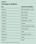
Getting rid of C pneumoniae in the laboratory is a straightforward process;the organism is susceptible to tetracyclines, macrolides, and quinolones,and resistant to sulfonamides (Table 2).25 In real life, treatmentdecisions are more complex. Most treatment studies have relied entirelyon diagnosis by serology, so microbiologic efficacy could not be assessed.Anecdotal reports suggest that prolonged courses, up to three weeks, oftetracyclines or erythromycin may be needed to eradicate C pneumoniae fromthe nasopharynxes of adults with flu-like illness and pharyngitis.9

Block and colleagues found that treatment with erythromycin suspensioneradicated C pneumoniae from 86% of culture-positive children with radiographicallyproven pneumonia. Clarithromycin suspension was essentially the same, eradicatingthe organism from 79%. This was surprising, since clarithromycin is moreactive in vitro than erythromycin and has superior pharmacokinetics andtissue penetration.5 All the children improved clinically despitepersistence of the organism in some.
We had a similar experience treating children with asthma. One of sixchildren who took clarithromycin continued to have positive cultures aftertwo 10-day courses of the drug; this child finally had a negative cultureafter treatment with erythromycin for three weeks.12 All sixculture-positive children treated with two weeks of erythromycin becameculture negative. The experience with azithromycin was very much the same.C pneumoniae was eradicated after treatment in 19 of 23 children (83%) withpneumonia who received azithromycin, in four of four who received amoxicillin-clavulanate,and in seven of seven who received erythromycin.7 All patientsimproved clinically despite persistence of the organism in some.
The success of amoxicillin-clavulanate was surprising but not totallyunexpected. Chlamydia species have penicillin-binding proteins, and amoxicillinhas activity in vitro, although it is variable. Amoxicillin is effectivefor treatment of genital C trachomatis infection in pregnant women and theCenters for Disease Control and Prevention recommends it for this indication.However, we need more data before we can recommend amoxicillin or amoxicillin-clavulanatefor treatment of C pneumoniae
infections.
On the basis of what data there are, we suggest the following regimensfor respiratory infection clinically suspected to be due to C pneumoniae:for children over 1 year of age,erythromycin suspension 50 mg/kg/d for 10to 14 days, clarithromycin suspension 15 mg/kg/d for 10 days, or azithromycinsuspension 10 mg/kg on day one followed by 5 mg/kg/d for four days. In childrenover 8 years of age, doxycycline 100 mg twice a day for 14 days can be used.Adult dosages can be used in adolescents. These dosage regimens are summarizedin Table 3.

Where does this leave us?
Most pediatricians are not going to be able to make a specific microbiologicdiagnosis of C pneumoniae infection in their patients. So what do you dowith the child presenting with an "atypical" community-acquiredpneumonia?
Most infectious disease experts would say, treat the child "as if"C pneumoniae were the causative organism. The assumption has a reasonableepidemiologic basis. Several multicenter studies indicate that C pneumoniaeand M pneumoniae are responsible for 10% to 40% of community-acquired pneumoniain children. Fortunately, both organisms are susceptible to the same antibiotics.For the time being, that's the best we can do.
THE AUTHOR is Professor of Pediatrics and Medicine and Director, Divisionof Pediatric Infectious Diseases, SUNY Health Science Center at Brooklyn,NY. She has research grants from Pfizer, Inc., and is a member of the Pfizerspeakers' bureau.
REFERENCES
1. Grayston JT, Campbell LA, Kuo CC, et al: A new respiratory tract pathogen:Chlamydia pneumoniae strain TWAR. J Infect Dis 1990;161:618
2. Falsey AR, Walsh EE: Transmission of Chlamydia pneumoniae.J InfectDis 1993;168:493
3. Yamazaki T, Nakada H, Sakurai N, et al: Transmission of Chlamydiapneumoniae in young children in a Japanese family. J Infect Dis 1990;162:1390
4. Ekman M-R, Grayston JT, Visakorpi R, et al: An epidemic of infectionsdue to Chlamydia pneumoniae in military conscripts. Clin Infect Dis 1993;17:420
5. Block S, Hedrick J, Hammerschlag MR, et al: Mycoplasma pneumoniaeand Chlamydia pneumoniae in community acquired pneumonia in children: Comparativesafety and efficacy of clarithromycin and erythromycin suspensions. PediatrInfect Dis J 1995;14:471
6. Jantos CA, Wienpahl B, Schiefer HG, et al: Infection with Chlamydiapneumoniae in infants and children with respiratory tract disease. PediatrInfect Dis J 1995;14:117
7. Harris JA, Kolokathis A, Campbell M, et al: Safety and efficacy ofazithromycin in the treatment of community acquired pneumonia in children.Pediatr Infect Dis J 1998;17:865
8.Shemer-Avni Y, Lieberman D: Chlamydia pneumoniae-induced ciliostasisin ciliated bronchial epithelial cells. J Infect Dis 1995;171:1274
9. Hammerschlag MR, Chirgwin K, Robin PM, et al: Persistent infectionwith C pneumoniae following acute respiratory illness. Clin Infect Dis 1992;14:178
10. Dean D, Roblin PM, Mandel L, et al: Molecular evaluation of serialisolates from patients with persistesnt C pneumoniae infections, in StephensRS, Byrne GI, Christiansen G, et al (eds): Chlamydial Infections: Proceedingsof the Ninth International Symposium on Human Chlamydial Infection. SanFrancisco, USCF, 1998, pp 219223
11. Hyman CL, Roblin PM, Gaydos CA, et al: Prevalence of asymptomaticnasopharyngeal carriage of C pneumoniae in subjectively healthy adults:Assessment by polymerase chain reaction-enzyme immunoassay and culture.Clin Infect Dis 1995;20:1174
12. Emre U, Roblin PM, Gelling M, et al: The association of C pneumoniaeinfection and reactive airway disease in children. Arch Pediatr AdolescMed 1994;148:727
13. Block S, Hammerschlag MR, Hedrick J, et al: C pneumoniae in acuteotitis media. Pediatr Infect Dis J 1997;16:858
14. Cook PJ, Davies P, Tunnicliffe W, et al: C pneumoniae and asthma.Thorax 1998;53:254
15. Emre U, Sokolovskaya N, Roblin PM, et al. Detection of anti-Cpneumoniae-IgEin children with reactive airway disease. J Infect Dis 1995;172:265
16. Tsumura N, Emre U, Roblin P, Hammerschlag MR: The effect of hydrocortisonesuccinate on the growth of C pneumoniae in vitro. J Clin Microbiol 1996;34:2379
17. Malinverni R, Kuo CC, Campbell LA, et al: Reactivation of C pneumoniaelung infection in mice by cortisone. J Infect Dis 1995;172:593
18. Miller ST, Hammerschlag MR, Chirgwin K, et al: The role of C pneumoniaein acute chest syndrome of sickle cell disease. J Pediatr 1991;118:30
19. Emre U, Bernius M, Roblin PM, et al: C pneumoniae infection in patientswith cystic fibrosis. Clin Infect Dis 1996;22:819
20. Tizer KB, Roblin PM, Gelling M, et al: C pneumoniae as a cause ofrespiratory illness in children with HIV infection. Pediatr Res 1998;43:159A
21. Montalban GS, Roblin PM, Hammerschlag MR: Performance of three commerciallyavailable monoclonal reagents for confirmation ofC pneumoniae in cell culture.J Clin Microbiol1994;32:1406
22. Kutlin A, Roblin P, Hammerschlag MR: Antibody response to C pneumoniaeinfection in children with respiratory illness. J Infect Dis 1998;177:720
23. Gaydos CA, Roblin PM, Hammerschlag MR, et al: Diagnostic utilityof PCREIA, culture and serology for detection of C pneumoniae in symptomaticand asymptomatic patients. J Clin Microbiol1994;32:903
24. Jantos CA, Roggendorf R, Wuppermann FN, et al: Rapid detection ofC pneumoniae by PCRenzyme immunoassay. J Clin Microbiol 1998;36:1890
25. Hammerschlag MR: Antimicrobial susceptibility and therapy of infectionsdue toC pneumoniae. Antimicrob Agents Chemother 1994;38:1873


