A Collage of Genital Lesions, Part 1
A discussion of three types of Genital Lesions: Phimosis, Paraphimosis, and Hypospadias.
A discussion of three types of genital lesions:

Phimosis
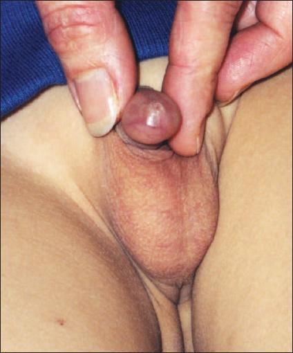
A 5-year-old boy presented with "ballooning" of the foreskin whenever he voided. He had a previous urinary tract infection and 2 episodes of balanoposthitis. On examination, the preputial opening was minute and the foreskin non-retractable.
Phimosis, the inability to pull back the foreskin, may result from narrowing or scarring of the preputial opening. Phimosis is physiological at birth and slowly resolves during infancy; by age 3 years, 90% of boys have a retractable foreskin. Phimosis might persist or develop consequent to forceful retraction of the foreskin, episodes of balanoposthitis, or other causes of foreskin inflammation. Ballooning of the foreskin when the child voids suggests relative obstruction at the preputial opening.
Treatment consists of careful attention to genital hygiene, gentle retraction of the foreskin, and the topical application of a hydrocortisone (eg, 0.05% betamethasone) cream twice a day to the foreskin edge and preputial opening. Attempts at forceful retraction and stretching may lead to pain, tearing of the foreskin meatus, bleeding, and scarring; such practice should be discouraged.
Circumcision should be considered if topical treatment with hydrocortisone cream is not successful; if the child experiences recurrent episodes of balanoposthitis; or if there is preputial scarring, balanitis xerotica obliterans, or urethral obstruction.
FOR MORE INFORMATION:
? Webster TM, Leonard MP. Topical steroid therapy for phimosis.
Can J Urol
. 2002;9:1492-1495. ? Zampieri N, Corroppolo M, Camoglio S, et al. Phimosis: stretching methods with or without application of topical steroids?
J Pediatr
. 2005;147:705-706.
A discussion of three types of genital lesions:

Paraphimosis
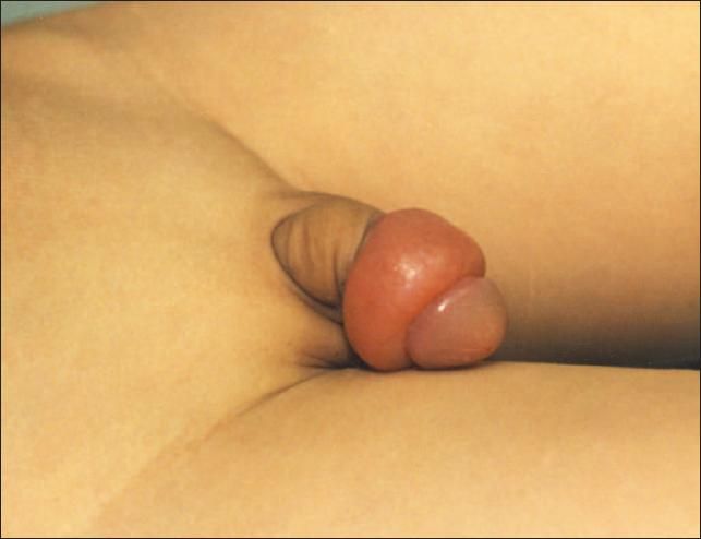
A 4-year-old boy presented 2 hours after his penis had become painful and swollen. There was no history of trauma, but the boy had been touching his penis just before the incident.
Paraphimosis develops when a phimotic foreskin is retracted past the coronal sulcus and becomes trapped. Lymphatic and venous stasis develops, with resultant pain and edema of the foreskin. The constricting band proximal to the swelling is pathognomonic.
Paraphimosis is a medical emergency. If the incarcerated prepuce is not released early, it may lead to arterial compromise and necrosis of the tip of the penis. Treatment consists of manual reduction of the foreskin with proper lubrication. Reduction of the foreskin can be achieved by applying direct pressure to the glans and placing distal traction on the foreskin to push the phimotic ring past the coronal sulcus. Infiltration of the edematous foreskin with hyaluronidase may facilitate decompression. If the foreskin cannot be reduced forward, a dorsal slit of the tight preputial ring might be required. A formal circumcision should be performed at a later date.
FOR MORE INFORMATION:
? Leung AK, Wong AL. Pediatric genital lesions.
Consultant for Pediatricians
. 2003;2:122-130.
A discussion of three types of genital lesions:

Hypospadias
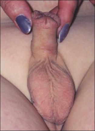
This 2-year-old boy was born with the external urethral meatus on the ventral surface of his penis.
The child had been born at 32 weeks to a 25-year-old gravida 2 para 1 mother. His birth weight was 1820 g and length was 38 cm. The mother's health was unremarkable during the pregnancy. There was no family history of a similar disorder. The boy had a weak urinary stream that was deflected downwards and splayed.
Hypospadias, the most common congenital anomaly of the penis, is characterized by a urethral meatus that is ectopically located proximal to the normal location and on the ventral aspect of the penis. The incidence is 0.4 to 8.2 per 1000 live male births. The condition is more common in white persons, least common among Hispanics, and of intermediate incidence in African Americans.
The cause is multifactorial. In most cases, the hypospadias develops sporadically without an obvious underlying cause. In general, the more severe the hypospadias, the more likely an underlying cause can be found. Defects in testosterone production by the testes and adrenal glands, failure of conversion of testosterone to dihydrotestosterone, deficient numbers of androgen receptors in the penis, or reduced binding of dihydrotestosterone to the androgen receptors can all result in hypospadias.
Intrauterine growth retardation and low birth weight are risk factors. A high familial incidence of hypospadias is observed and a polygenic predisposition is likely. Hypospadias has been found in various chromosomal aberrations, such as 4p-, 18q-, paracentric inversion of chromosome 14, and Klinefelter syndrome.
The meatal position can be classified as anterior or distal (glandular, coronal, subcoronal), middle (midpenile), or posterior or proximal (posterior penile, penoscrotal, scrotal, perineal). The subcoronal position is the most common. Characteristically, the foreskin on the ventral surface is thin or absent while the foreskin on the dorsal side is abundant; this gives the appearance of a dorsal hood. Chordee is more commonly associated with proximal hypospadias. Chordee becomes more apparent with penile erection and might only be noticeable during an erection.
Approximately 8% to 10% of boys with hypospadias have cryptorchidism and 9% to 15% have associated inguinal hernia. Urinary tract anomalies occur in 1% to 5% of cases with isolated anterior and posterior hypospadias, respectively.1 Complications of untreated hypospadias include deformity of the urinary stream, inability to void in the standing position, sexual dysfunction, and infertility.
Laboratory tests are usually not indicated for isolated anterior or middle hypospadias. Karyotyping should be performed in patients with cryptorchidism or ambiguous genitalia. A voiding cystourethrogram should be considered in patients with proximal hypospadias.
Many procedures have been designed for the repair of hypospadias, including meatal advancement-glanuloplasty, the glans approximation procedure, and tubularization following incision of the urethral plate. The ideal age for surgical repair in a healthy child is approximately 6 to 12 months. Most cases can be repaired in a single operation.
Circumcision is contraindicated in patients with hypospadias because the foreskin is essential for hypospadias repair.
FOR MORE INFORMATION:
? Baskin LS. Hypospadias.
Adv Exp Med Biol.
2004;545:3-22.
? Leung AK, Robson WL. Hypospadias: an update.
Asian J Androl
. 2007;9:16-22.
References:
- Baskin LS. Hypospadias. Adv Exp Med Biol. 2004;545:3-22.