Do You Recognize These Eye Disorders?
The parents of this child are concerned that her eyes are “crossed.” Is this condition cause for concern?
Case 1:
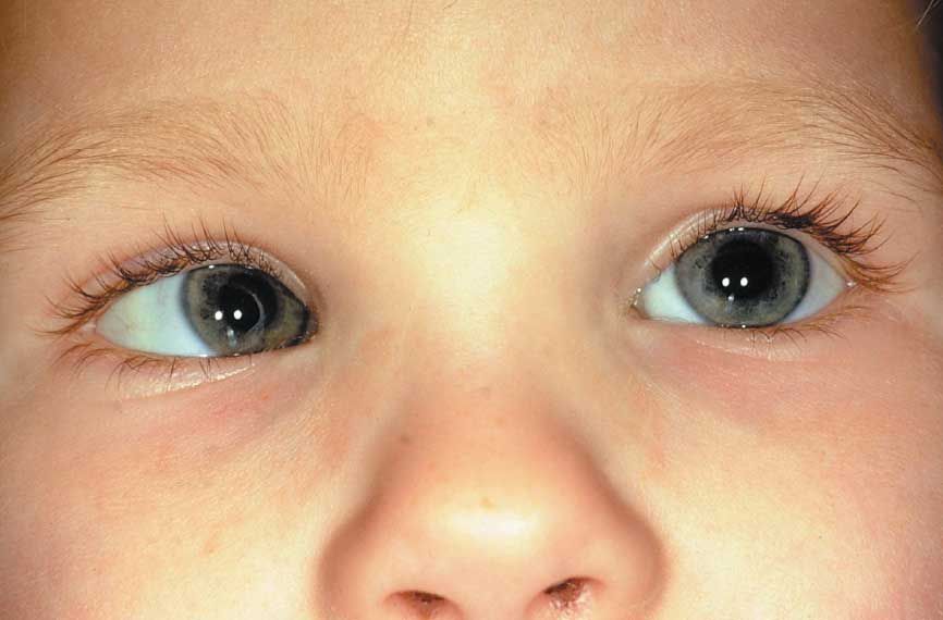
The parents of this child are concerned that her eyes are “crossed.” Is this condition cause for concern?

Case 1: Patients with strabismus display ocular deviation in an inward (esotropia, A), outward (exotropia, B), upward (hypertropia), or downward (hypotropia) direction. A head tilt may also be evident as the child attempts to improve vision.
Strabismus must be distinguished from pseudostrabismus. Such features as a wide nasal bridge, large epicanthal folds, and widely or closely spaced eyes may give the illusion of crossed eyes; however, the eyes are not truly misaligned.
Ask whether the deviation is constant or intermittent, whether recent trauma has occurred, and whether there is a family history of strabismus. A neurologic and ophthalmologic examination must be complete: examine visual acuity, pupil reactivity, eyelid position, and extraocular movements. Note the presence of a head tilt.
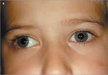
The corneal light refection test (Hirschberg method) is the initial screen for strabismus. To conduct the test, hold a small toy several feet in front of the child’s face, and hold a penlight next to the toy. If alignment is normal, the light reflection will be positioned at the same point in each eye. If there is misalignment, the light will be deflected in the affected eye.
The corneal light reflection test is used in conjunction with the single cover test and the alternate cover test. During the single cover test, ask the patient to fixate on a target while you cover one eye and observe the opposite eye for movement. If the uncovered eye shifts to refixate on the target, strabismus is present: refer the patient to an ophthalmologist.1,2 Examine both eyes in this manner.
For the alternate cover test, have the child fixate on a target while you cover one eye for a few seconds and then quickly move the cover to the other eye. Watch the uncovered eye closely for movement, which can indicate a latent strabismus (phoria)1,2-a finding that may not be obvious on initial examination. The amount of movement may increase as the cover is rapidly alternated.
The pathophysiology of the most common forms of childhood strabismus is not fully understood. In the visually immature newborn, strabismus occurs sporadically and resolves within the first few months of life as binocular vision improves.
Infants with strabismus may have subtle abnormalities in both motor and binocular sensory functions. Dysconjugate eye movements may be seen in normal infants up to 4 months of age. Esodeviations are the most common type of strabismus in the pediatric population.2 Congenital esotropia is a large angle esotropia that presents from birth to 6 months of age and may be associated with vertical deviations as well as abnormal saccadic eye movements and nystagmus. Accommodative esotropia presents later (6 months to 7 years) and is caused by abnormalities in the accommodative convergence reflex that is needed to look at near objects.2 This is often the result of uncorrected hyperopia or the inability of the eyes to diverge.
Acute-onset strabismus may result from many intracranial processes (hemorrhage, infection, tumor) and other nervous system disorders, such as the Miller Fisher variant of Guillain-Barr syndrome, ventriculoperitoneal shunt failure, lead poisoning, and myasthenia gravis.
The treatment for strabismus depends on its cause. Some congenital forms of esotropia must be treated surgically if eyeglasses or cycloplegic eye drops cannot correct the condition. Treatment for exotropia focuses first on the underlying cause of the misalignment- whether it is from refractive error or from other causes of amblyopia. After the amblyopia has been adequately treated, surgical intervention is then indicated if the strabismus persists. To treat the amblyopia, the initial management involves occlusion or penalization of the preferred eye with the use of prescription glasses, ophthalmic drops that manipulate accommodation, and occlusive patches. These medical therapies are also used to correct alignment. However, surgical intervention is sometimes necessary to improve alignment and involves shortening and repositioning the insertion of the rectus muscle. Patients with exotropia or esotropia deserve prompt ophthalmologic referral.1,2
Case 2:
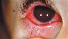
For 2 days, this 2-year-old boy has had red eyes, a large amount of drainage from his eyes, and matted eyelashes.
What does this condition look like to you?

Case 2: Conjunctival injection can be caused by a variety of conditions, including infections (viral and bacterial), allergies, trauma, and foreign bodies. In children, however, bacterial infections are the most common cause of conjunctivitis: the most frequent pathogens are Haemophilus influenzae and Streptococcus pneumoniae.1 Recently, there has been an increase in the number of cases of conjunctivitis caused by gram-positive organisms. This child has acute bacterial conjunctivitis.
Other conditions that figure into the differential diagnosis of red eyes and drainage are blepharitis and pre-septal and orbital cellulitis.
Patients with conjunctivitis present with conjunctival injection and drainage. The drainage is mucopurulent if the infection is bacterial and watery, or mucoid if the infection is viral. Many patients-especially those with bacterial conjunctivitis-have matted eyelashes. Bacterial and allergic conjunctivitis are usually bilateral, while viral conjunctivitis is usually unilateral. In many patients with viral conjunctivitis, there is an associated upper viral infection. About 20% of patients with viral conjunctivitis have preauricular or submandibular lymphadenopathy.2 Allergic conjunctivitis is associated with itching.
Culture of the eye drainage is not usually required because results are unavailable for several days. Also, with topical broad-spectrum antibiotics, culture results do not change therapy. Culture is indicated, however, in neonates with conjunctivitis that does not respond to topical antibiotics and also in patients with suspected chlamydial infection.
Topical antibiotics shorten the duration of bacterial conjunctivitis and eradicate the bacteria from the conjunctiva. A child can attend school once therapy is started; the treatment also reduces the spread of infection in daycare centers and schools. A fourth-generation fluoroquinolone-specifically moxifloxacin or gatifloxacin-is recommended. Moxifloxacin is preferred in this setting because its pH is close to the pH of natural tears; moreover, the agent is preservative-free and needs to be used less frequently (three times a day) than do other fluoroquinolones.3Case 3:
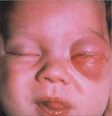
This extremely irritable infant presented with red, swollen eyelids and a temperature of 39ºC (102.2ºF). CT scan of the orbits revealed inflammatory changes in the post-septal area. What is the likely diagnosis?

Case 3: The inflammatory changes in the post-septal area are consistent with orbital (or post-septal) cellulitis.
Periorbital inflammation has many causes besides cellulitis, including insect bites, blepharitis, and conjunctivitis. In addition to infections, any infiltrative process (ie, a malignancy) that disrupts the post-septal space can lead to an inflamed periorbital area.
Orbital cellulitis arises secondary to spread of infection from structures adjacent to the walls of the orbit-especially from the ethmoidal sinuses. Trauma and the spread of infection from the oral cavity also may lead to orbital cellulitis. The valveless venous system in this area adds to its vulnerability to infection from adjacent structures. Ophthalmic vein and cavernous venous thrombosis are potential complications.
The responsible bacterial agents are the same as those that cause pre-septal, sinus, and dental infections: Streptococcus pneumoniae, Haemophilus influenzae, Moraxella catarrhalis, Staphylococcus aureus, and anaerobes.1-3
A thorough history and physical examination that focuses on the structures of the head is the initial step in the evaluation. An eye examination and evaluation for pain with eye movement, proptosis, and extraocular muscle impairment are essential to provide evidence of involvement of the post-septal space. Obvious periorbital inflammation with eye pain, fever, headache, proptosis, and restricted extraocular movements strongly suggests a diagnosis of orbital cellulitis.
A contrast-enhanced CT scan of the orbit is the test of choice for diagnosing orbital infections.4
Because orbital cellulitis can have devastating complications (intracranial sequelae, vision loss, and even death), aggressive management is required. Consultation with an ophthalmologist and otolaryngologist is needed to establish the appropriate treatment plan for a particular patient (intravenous antibiotics alone or with surgery) and for close monitoring of vision, extraocular muscle function, and degree of proptosis.
Case 4:
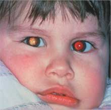
This infant was found to have an asymmetric red reflex during a routine checkup.
What are the possible causes, and what is the most likely diagnosis?

Case 4: The asymmetric red reflex is a manifestation of retinoblastoma-the most common intraocular malignancy of childhood-which typically presents as leukokoria. 1 A parent may bring this condition to the pediatrician’s attention, or it may be detected during a physical examination in the first 1 to 2 years of life. Strabismus or visual disturbance may also be a presenting complaint. A red, painful eye secondary to glaucoma, orbital cellulitis, vitreous hemorrhage, and microphthalmos are rare presenting findings in retinoblastoma.1
Leukokoria can be caused by many entities- such as congenital cataracts, toxocariasis, retinal detachment, and Coats disease (exudative vasculopathy of retinal vessels). Tumors that can occupy the same space include hemangiomas, melanomas, and teratomas. A thorough history (including questions about a family history of retinoblastoma) and an eye examination that includes a red reflex test are essential.
To test the red reflex, focus the ophthalmoscope on each pupil at 12 to 18 inches from the patient in a darkened room.2 The red reflex should be bright reddish yellow or, in dark-skinned persons, light gray. Worrisome findings that warrant immediate referral to a pediatric ophthalmologist include the lack of a red reflex; or a red reflex that is white, blunted, or that displays dark spots.2 The ophthalmologist may obtain a biopsy of the tumor. Ultrasonography and CT scans of the orbits and brain are needed to further delineate the disease burden. Other intracranial tumors (eg, a pineal gland tumor) may occur with retinoblastoma.
Retinoblastoma develops in the immature retina; the cell of origin is neuronal. There are familial and sporadic cases. Familial retinoblastoma is considered an autosomal dominant inherited cancer syndrome. Carriers of a mutant Rb tumor suppressor gene are at great risk for bilateral retinoblastoma and are also at increased risk for suffering from a secondary cancer-particularly osteogenic sarcoma.3
Retinoblastoma is lifethreatening and must be treated aggressively. When choosing therapy, the extent of disease must be weighed against the potential toxicity of available treatments. Advanced retinoblastoma mandates enucleation, but chemotherapy and local therapies may suffice for those who have minimal to moderate disease. Family members of the affected child should also be screened for retinoblastoma.
References:
REFERENCES for Case 1:
1. Bradford CA. Amblyopia and strabismus. In: Bradford CA, ed.
Basic Ophthalmology for Medical Students and Primary Care Residents
. 7th ed. San Francisco: American Academy of Ophthalmology; 1999:94-106.
2. Simon JW, ed.
Pediatric Ophthalmology and Strabismus
. San Francisco: American Academy of Ophthalmology; 2002:7-124.
REFERENCES for Case 2:
1. Ambati BK, Ambati J, Azar N, et al. Periorbital and orbital cellulitis before and after the advent of Haemophilus influenzae type B vaccination.
Ophthalmology
. 2000;107:1450-1453.
2. Schwartz GR, Wright SW. Changing bacteriology of periorbital cellulitis.
Ann Emerg Med
. 1996;28:617-620.
3. Bekibele CO, Onabanjo OA. Orbital cellulitis: a review of 21 cases from Ibadan, Nigeria.
Int J Clin Pract
. 2003;57:14-16.
4. Mafee MF, Mafee RF, Malik M, Pierce J. Medical imaging in pediatric ophthalmology.
Pediatr Clin North Am
. 2003;50:259-286.
REFERENCES for Case 3:
1. Ambati BK, Ambati J, Azar N, et al. Periorbital and orbital cellulitis before and after the advent of Haemophilus influenzae type B vaccination.
Ophthalmology
. 2000;107:1450-1453.
2. Schwartz GR, Wright SW. Changing bacteriology of periorbital cellulitis.
Ann Emerg Med
. 1996;28:617-620.
3. Bekibele CO, Onabanjo OA. Orbital cellulitis: a review of 21 cases from Ibadan, Nigeria.
Int J Clin Pract
. 2003;57:14-16.
4. Mafee MF, Mafee RF, Malik M, Pierce J. Medical imaging in pediatric ophthalmology.
Pediatr Clin North Am
. 2003;50:259-286.
REFERENCES for Case 4:
1. Abramson DH, Frank CM, Susman M, et al. Presenting signs of retinoblastoma.
J Pediatr
. 1998;132:505-508.
2. Committee on Practice and Ambulatory Medicine, Section on Ophthalmology; American Association of Certified Orthoptists; American Association for Pediatric Ophthalmology and Strabismus; American Academy of Ophthalmology. Eye examination in infants, children, and young adults by pediatricians.
Pediatrics
. 2003;111:902-907.
3. Kumar V, Abbas AK, Fausto N, Robbins SL. Neoplasia. In: Robbins & Cotran Pathologic Basis of Disease. 7th ed. Philadelphia:
Elsevier Saunders
; 2005:284.