Instrument-based vision screening: Update and review
Insurance companies are now beginning to compensate pediatricians for performing photoscreening, billed under Current Procedural Terminology (CPT) code 99174. We applaud the efforts of the many pediatricians, pediatric ophthalmologists, and state chapters of the AAP who have aggressively petitioned insurance companies to cover this important service for our patients. -Andrew J Schuman, MD, Section Editor
In late 2012, the American Academy of Pediatrics (AAP) Section on Ophthalmology and Committee on Practice and Ambulatory Medicine joined the American Academy of Ophthalmology, the American Association for Pediatric Ophthalmology and Strabismus (AAPOS), and the American Association of Certified Orthoptists in issuing a policy statement endorsing the use of instrument-based vision screening in the pediatric population.1 This was the subject of the inaugural article in the Peds v2.0 series in January 2013 (Contemp Pediatr. 2013:30(1):41-44). We now revisit the topic because insurance companies are beginning to compensate pediatricians for performing photoscreening, billed under Current Procedural Terminology (CPT) code 99174. We applaud the efforts of the many pediatricians, pediatric ophthalmologists, and state chapters of the AAP who have aggressively petitioned insurance companies to cover this important service for our patients. -Andrew J Schuman, MD, Section Editor
Amblyopia, defined as poor vision caused by abnormal development of visual areas of the brain, occurs in as many as 2% to 4% of children.2 It is associated with complete or partial lack of clear visual input to 1 eye (unilateral/anisometropic refractive amblyopia), or, less often, to both eyes (bilateral refractive amblyopia), or to conflicting visual inputs to the 2 eyes (strabismic amblyopia). Less-common causes
include ptosis, congenital cataract, and corneal injury or dystrophy. According to the US Preventive Services Task Force (USPSTF), amblyopia is regarded as a disease of childhood; however, its effects are irreversible if left untreated, and it is the most common cause of monocular vision loss among adults aged 20 to 70 years.3
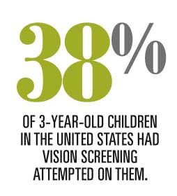
Unfortunately only a minority of young children are screened for this disabling condition. One study reported that in a sample of 102 private pediatric practices in the United States, vision screening was attempted on only 38% of 3-year-old children and 81% of 5-year-old children.4 The study also showed that only 21% of children failing the AAP vision screening guidelines were referred for a professional eye examination.
Vision screening in pediatric practice
Vision and amblyopia screening should be viewed as a continual process beginning in mid-infancy and repeated annually throughout early childhood.5 Vision screening for infants, toddlers, and preschool children is traditionally performed by primary care physicians and nurses, using a rechargeable, battery-powered ophthalmoscope to test pupil position, equality, size, steadiness, and reaction to bright light; extraocular muscle function; ocular deviation during a cover-uncover test; and presence of unequal red reflexes in a darkened room.
Many of the major risk factors for amblyopia are poorly detected during a traditional vision screening examination. Vision acuity testing in children aged younger than 3 years in a medical office can be challenging, because few children this age can be screened with a vision chart. From ages 3 to 5 years, screening is possible with Snellen charts, Tumbling E charts, or picture tests such as Allen Visual Acuity Cards, but this is time consuming and can lead to inconsistent or erroneous results.
Amblyopia: the problem
Amblyopia remains treatable until age 60 months, with rapid decline of effective treatment after age 5 years.5,6 The goal of vision screening in infants and young children, therefore, must be the early detection of high severity (magnitude) amblyopia risk factors (ARFs), including moderate or severe astigmatism, anisometropic myopia, high hyperopia, severe strabismus, and opacities in the visual axis, including retinoblastoma or other ocular entities that cause opacities that interfere with transmission of light to and from the retina (Table 13,7). Any media opacity (including the retina) greater than 1 mm in size is potentially amblyogenic and should be detected with photoscreening.
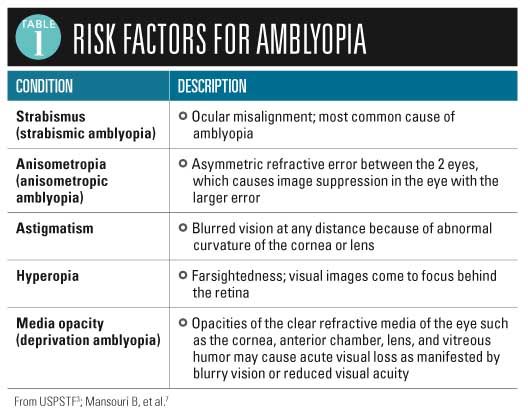
The relationship between refractive error and the likelihood of the development of amblyopia depends on the child’s age, the magnitude of blurred vision, and other factors.5 For children aged 3 years and younger, the prevalence of amblyopia correlates with the severity of anisometropia (unequal refractive power between each eye).8 In this age group, high-magnitude amblyopia risk factors increase the likelihood of overt amblyopia. Many children with mild amblyopia from anisometropic amblyopia or strabismic amblyopia improve with spectacle (eyeglasses) treatment alone.9 Treatment options for children in whom spectacles are not appropriate or are not effective include patching the better-seeing eye or using atropine drops to blur vision in the better-seeing eye.
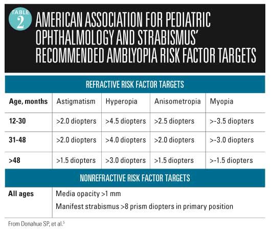
In 2013, the AAPOS Vision Screening Committee revised its criteria for instrument-based vision screening because “many children with low-magnitude ARFs do not develop amblyopia, and those who do often respond to spectacles alone.”5 They noted that referral criteria for vision photoscreening instruments should have high specificity for ARFs in young children (to minimize false positive referrals) and high sensitivity to detect amblyopia risk factors in older children (when children are approaching an age when treatment becomes less effective).
See Table 2 for the current age-related thresholds recommended by the AAPOS. Note that the detection of higher magnitude anisometropia (>3.0 diopters), even in children in the 31- to 48-month age group, should be highly sensitive because it is associated with amblyopia that continues to deepen over time.
Visually significant media opacities such as cataracts, infantile glaucoma, persistent hyperplastic primary vitreous, and retinoblastoma must be detected accurately at all ages.7
Benefits of photoscreening
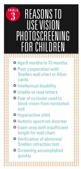
In primary care pediatric and family medicine offices, photoscreening for high refractive errors and other ARFs is recommended for infants, toddlers, and preschool-aged children. Even in primary grades, such technology offers advantages over traditional Snellen wall charts or Allen cards because it is less time consuming, more efficient, and provides much more specific information (Table 3).1 In the Vision in Preschoolers Study, published in 2004, it was found that visual acuity testing (using eye charts) of more than 2,500 preschool children had a 77% sensitivity for detecting conditions associated with amblyopia, while photoscreener devices had a sensitivity of 81% to 88%.10 Note that the use of photoscreeners not only improves detection of eye pathology but also does so in a fraction of the time required to perform testing with eye charts.
How to photoscreen
Regardless of the device used to screen patients, testing should be done in a darkened room with lights off and, if necessary, window shades drawn. If the room is too dark, the room door may be opened a small amount. The child’s pupils should be level. Head tilting produces off-center pupils that can produce false positive results for refraction and astigmatism problems. The child’s pupil size must be at least 4 mm in diameter to aim the light beam into the plane of the visual axis. If the subject’s pupil size is too small for the light beam to penetrate well, the instrument will state: “pupils too small.” Children from many Asian countries have epicanthic folds or partial ptosis, which require them to open their eyelids as wide as possible for the light beam to penetrate into their pupils.
Photoscreening devices
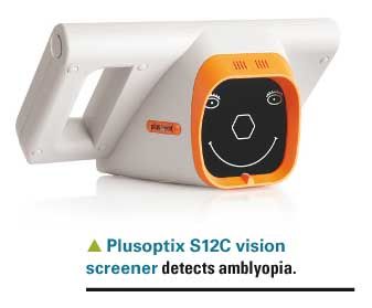
Pediatricians interested in incorporating photoscreening technology into routine practice have a number of devices to choose from. Dr. Schuman detailed several of these devices in the January 2013 article. We provide a brief description of popular photoscreeners here along with some new devices that either are presently available or will be in the near future. Note that many insurance companies are now covering photoscreening under the CPT code 99174 (“ocular photoscreening with interpretation and report, bilateral”), paying usually $25 to $30 per screen.
Plusoptix (Atlanta, Georgia), a German company, introduced its 4th-generation vision screener last year-the portable Plusoptix model S12C. Patient information is inputted with a touch-screen interface, and then pushing a trigger button activates the camera. The device produces a warbling sound to attract the child’s attention and gaze toward the smiling face displayed on the patient side of the device.
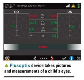
The child’s eyes are positioned in a white rectangle on the viewing screen and moved until a green line is drawn between the pupils. Once positioned, the device takes 36 pictures of both eyes and performs measurements regarding pupil sizes, corneal reflexes, and refraction, which are displayed on the screen along with an indication whether the child passed the screening.
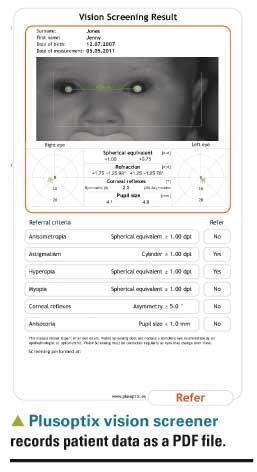
The device records the patient data as a pdf file on a secure digital card that can be transferred to a computer to print the result for incorporation into the patient record. It connects wirelessly to a Zebra printer to print an abbreviated version of the results. According to Plusoptix, the device can detect the amblyopia with a sensitivity as high as 92% and specificity as high as 88%. The S12C device sells for $7,385 each. A smaller version of the Plusoptix portable device, model S12R, doesn’t have the wireless printing capability or data storage capability of the model C, but is priced at $5,875.
Spot Vision Screener from PediaVision (Lake Mary, Florida) is a photoscreener that is battery powered, lightweight (2.5 pounds), and features a portable infrared camera that, like the Plusoptix S12C, combines autorefraction and video retinoscopy. The rechargeable battery is designed to supply sufficient power for an entire day of screening children. Spot makes 23 images of the retina by
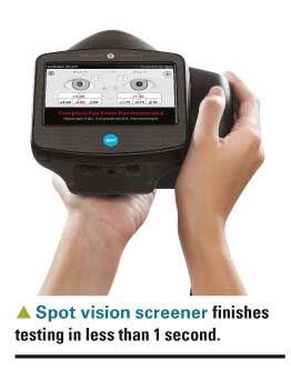
shooting infrared light through a 4-mm or greater pupillary aperture. The light beam is directed to the retina but may be blocked by lesions in the cornea, lens, vitreous, and retina itself. In the absence of opacified lesions in the visual axis, the light beam is reflected off the retina and returns to the instrument. A software program analyzes the result and compares it to internal norms that can be modified for the child’s age.
Physicians familiar with digital cameras find the Spot easy to use. After inputting identifying information, one aims the device toward the child and positions the child’s face on a display. The device indicates whether you are too close or too far from the subject and then initiates the screening sequence, which is completed in less than a second. The screen displays the pupillary size, distance, alignment, and complete refraction information for both eyes and indicates whether the child needs to be referred to a pediatric ophthalmologist. Additionally, the device can connect to a wireless printer to facilitate printing a complete report for parents or for inclusion in the medical record. The Spot Vision Screener sells for $7,495. Those pediatricians who purchased the Spot more than 9 months ago need to reset the device’s threshold to those listed in Table 2 to avoid overreferrals to ophthalmologists.5
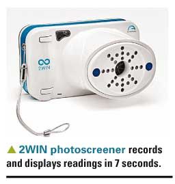
The 2WIN binocular refractometer and photoscreener is new to the American market, produced by an Italian company called Adaptica (Padova, Italy). It is comparable in size to the small Plusoptix S12R. According to the manufacturer, 2WIN has a rechargeable battery; records and displays readings in as little as 7 seconds; and has wireless and infrared printer connectivity. The device is priced at $6,950.
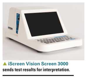
The iScreen Vision Screen 3000 from iScreen Vision Inc (Cordova, Tennessee), is yet another photoscreener that takes pictures of the pupillary and red reflex to screen for amblyopia. In contrast to the automatic computer analysis of the photos of the red reflex performed by the Plusoptix and PediaVision devices, the iScreen Vision Screen 3000 connects by a network cable to your office network and transfers each patient screening test to a “professional” who interprets the test (with computer assistance) and provides information regarding whether the patient is at risk for amblyopia and needs referral to a pediatric ophthalmologist. Results are transmitted to the office e-mail within minutes of receipt, so the parents can be apprised of the results before the visit is done. The iScreen Vision Screen 3000 sells for $4,200, and each test costs $6.
New screening technologies coming soon
If there is a limitation to photoscreeners, it is that they can still miss a child with amblyopia, and there is a substantial overreferral rate associated with these devices. Hence, it is important that screening is
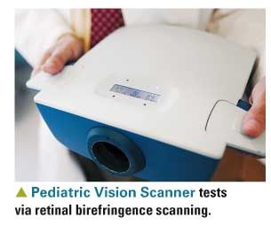
performed yearly at well-child checks, and when a child is referred to an ophthalmologist, parents must be reminded that there is a possibility that no pathology will be discovered. David Hunter, MD, chief of ophthalmology at Boston Children’s Hospital, has developed a new device called the Pediatric Vision Scanner (PVS) that uses polarized laser light to test eye orientation at the retinal level to detect amblyopia in children with sensitivities and specificities much higher than those associated with photoscreeners. The new technology is called “retinal birefringence scanning,” and Hunter has formed a company called REBIScan to commercialize its use. The PVS is currently being evaluated by the US Food and Drug Administration and, following approval, will be available for use by pediatricians this year.
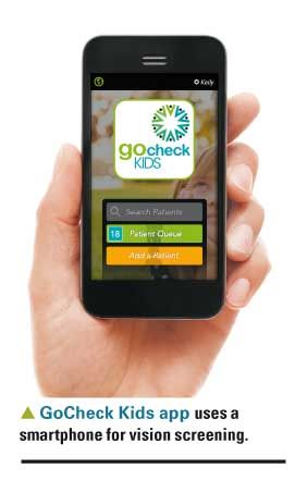
Lastly, a new company called Gobiquity (Aliso Viejo, California) will soon introduce an iPhone application called GoCheck Kids that uses the camera built into the smartphone to photograph a child’s eyes. The application will subsequently analyze the photograph in much the same way as photoscreeners do, generating a report that will indicate whether the child passes or needs referral to a pediatric ophthalmologist. Initial data indicate that the GoCheck Kids application produces results comparable to those of currently available photoscreeners. The cost of the application and pricing model for screening has not yet been determined as of this writing.
Conclusion
Author Richard Schwartz, MD, has been using the Spot instrument in his practice since May 2013, and has been able to screen young and often uncooperative children for refractive errors or amblyopia risk factors. It has paid for itself in this time and has detected several children with significant refractive errors as well as a cataract and posterior vitreous disease. He reports that insurance companies are reimbursing for photoscreening in Virginia.
Author Andrew Schuman, MD, has trialed both the Plusoptix S12C and the Spot and has found them easy to use-both providing readings in seconds. He notes that parents are very impressed that their children can be screened so easily, and using either device is a huge time-saver in his pediatric office. In his state of New Hampshire, all insurance carriers, including Medicaid, are currently reimbursing for photoscreening. He advises that before you purchase, you contact your insurance companies to see if they will reimburse for CPT code 99174.
Practices should challenge rejected claims by providing copies of the new AAP policy and/or the USPSTF policy statement, and when necessary ask the state chapter of the AAP to petition insurance companies to cover this important service as well.
REFERENCES
1. Miller JM, Lessin HR; American Academy of Pediatrics Section on Ophthalmology; Committee on Practice and Ambulatory Medicine; American Academy of Ophthalmology; American Association for Pediatric Ophthalmology and Strabismus; American Association of Certified Orthoptists. Instrument-based pediatric vision screening policy statement. Pediatrics. 2012;130(5):983-986.
2. Friedman DS, Repka MX, Katz J, et al. Prevalence of amblyopia and strabismus in white and African American children aged 6 through 71 months the Baltimore Pediatric Eye Disease Study. Ophthalmology. 2009;116(11):2128-2134.e1-2.
3. US Preventive Services Task Force. Vision screening for children 1 to 5 years of age: US Preventive Services Task Force Recommendation Statement. Pediatrics. 2011;127(2);340-346.
4. Wasserman RC, Croft CA, Brotherton SE. Preschool vision screening in pediatric practice: a study from the Pediatric Research in Office Settings (PROS) Network. American Academy of Pediatrics. Pediatrics. 1992;89(5 pt 1):834-838. Erratum in: Pediatrics. 1992;90(6):1001.
5. Donahue SP, Arthur B, Neely DE, Arnold RW, Silbert D, Ruben JB: POS Vision Screening Committee. Guidelines for automated preschool vision screening: a 10-year, evidence-based update. J AAPOS. 2013:17(1):4-8.
6. Holmes JM, Lazar EL, Melia BM, et al; Pediatric Eye Disease Investigator Group. Effect of age on response to amblyopia treatment in children. Arch Ophthalmol. 2011;129(11):1451-1457.
7. Mansouri B, Stacy RC, Kruger J, Cestari DM. Deprivation amblyopia and congenital hereditary cataract. Semin Ophthalmol. 2013;28(5-6):321-326.
8. Donahue SP. Relationship between anisometropia, patient age, and the development of amblyopia. Am J Ophthalmol. 2006;142(1):132-140.
9. Cotter SA; Pediatric Eye Disease Investigator Group. Edwards AR, Wallace DK, Beck RW, et al. Treatment of anisometropic amblyopia in children with refractive correction. Ophthalmology. 2006;113(6):895-903.
10. Schmidt P, Maguire M, Dobson V, et al; Vision in Preschoolers Study Group. Comparison of preschool vision screening tests as administered by licensed eye care professionals in the Vision in Preschoolers Study. Ophthalmology. 2004;111(4):637-650.
Alaska Blind Child Discovery brings vision screening to Alaska’s preschoolers
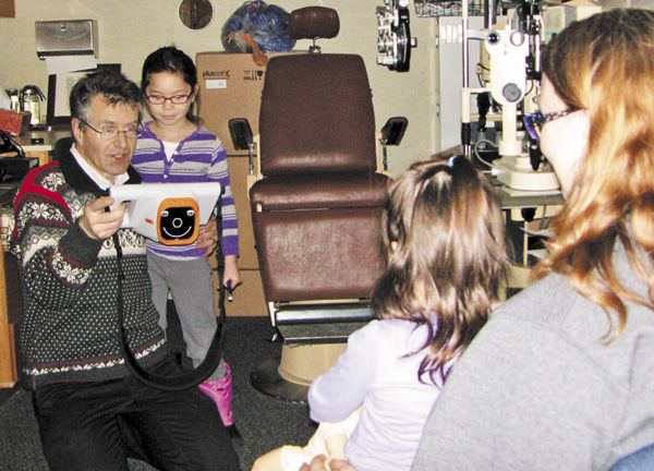
Dr. Arnold tests patients for amblyopia.There is no such thing as a “brief” conversation with Robert W. Arnold, MD, pediatric ophthalmologist, when the topic is preschool vision screening. He originated the Alaskan Blind Child Discovery (ABCD) program in his beloved state of Alaska in the early 1990s. Its goal was to test every young child in Alaska for amblyopia.
Arnold began with a handful of photoscreener devices that he provided to pediatricians, public health nurses, and ophthalmologists. Later, he gained philanthropic support provided by local Lions, Kiwanis, and Shriners organizations and launched vision screening clinics throughout the state.
Along the way, Arnold found the time to perform many investigations that helped refine and ultimately define the current state-of-the-art regarding vision screening for young children. He has published extensively and is a coauthor of the 2013 AAPOS guidelines for automated preschool vision screening.5
From 1996 through today, the ABCD project has screened over 60,000 children. The project’s website, www.abcd-vision.org/, has a wealth of information regarding pediatric vision screening that can assist pediatricians interested in starting their own vision programs. There also are numerous informative videos, with many demonstrating the photoscreeners discussed in this article.
Author supplied images.
DR SCHWARTZ is professor of pediatrics, Inova Children’s Hospital, Falls Church, Virginia, satellite of Virginia Commonwealth University School of Medicine, Richmond, and co-partner, Advanced Pediatrics, Vienna, Virginia. DR SCHUMAN is adjunct associate professor of pediatrics, Geisel School of Medicine at Dartmouth, Lebanon, New Hampshire, and section editor for Peds v2.0 and editorial advisory board member for Contemporary Pediatrics. DR WEI is on active staff, Department of Surgery, Section of Ophthalmology, Virginia Hospital Center, Arlington. The authors have nothing to disclose in regard to affiliations with or financial interests in any organizations that may have an interest in any part of this article.
Anger hurts your team’s performance and health, and yours too
October 25th 2024Anger in health care affects both patients and professionals with rising violence and negative health outcomes, but understanding its triggers and applying de-escalation techniques can help manage this pervasive issue.