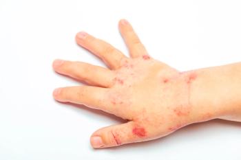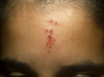
Painful lumps: Cause for concern?
Lumps in children that cause pain have six basic causes. Here's how to differentiate among them and determine which require further investigation.
Painful lumps: Cause for concern?
By Joanna M. Burch, MD, and William L. Weston, MD
Lumps in children that cause pain have six basic causes. Here's how to differentiate among them and determine which require further investigation.
Many conditions can cause a lump or mass in a child's skin. Most masses are asymptomatic. A few are characteristically or frequently tender, however. When confronted with a painful lump in a child's skin, careful palpation with the thumb and forefinger can determine whether the lesion is soft or firm, mobile or bound down, and multilocular or not (Figure 1). These characteristics, along with the color of the lesion, can help you distinguish the cause of the lump and decide what, if anything, to do about it.
Six most common causes of painful lumps
Six entities account for most painful lumps in children:
- calcifying epithelioma of Malherbe (pilomatricoma)
- angiolipoma
- neurofibroma
- dermatofibroma
- lymph node (enlarged)
- eccrine spiradenoma (less common than the others)
An easy way to remember these lumps is the mnemonic CANDLE (see the
Calcifying epitheliomas of Malherbe (pilomatricomas) are benign tumors of hair germ cells. They appear most often as solitary, tender nodules on the head, neck, and proximal extremities, although multiple lesions have been reported. Despite their origin in the hair matrix, very few are found on the scalp. It has been suggested that hair follicle density is a factor in their formationthe greater the density, the more likely the tumors are to developand the follicle density of the scalp is half that of the face even though the scalp has more hair follicles.1
Calcifying epitheliomas typically occur in the first 15 years of life. They vary in diameter from 0.5 cm to as large as 15 cm; most are between 0.5 and 1.5 cm. In rare cases, occurrence follows a familial pattern.2,3,4
Calcifying epitheliomas are dermal tumors, usually covered by normal or translucent, bluish skin. They calcify with time and tend, on physical examination, to be mobile, multiloculated, and rock-like (Figure 2). Malignant transformation is rare but has been reported in adults.5 Because the lesions do not regress spontaneously,4 excision is the treatment of choice.
Angiolipomas are benign fat cell tumors with proliferating blood vessels. They appear as tender, compressible, subcutaneous masses, most commonly on the upper outer arms of adolescent girls. The initial large study of angiolipomas found that most patients were 16 years of age or older and that the lesions appeared most often on the extremities and trunk.6 Masses are often 2 to 8 cm in diameter. The innervated vasculature of the tumors is believed to cause the pain. Treatment is surgical excision.
Neurofibromas are soft, fleshy papules ranging from skin-colored to dusky brown (Figure 3). On direct palpation perpendicular to the skin, they often display the so-called buttonhole sign; that is, the examiner can feel an opening just below the lesion into which the lesion can be indented.
Neurofibromas originate from cells of the nerve sheath. They can occur anywhere on the skin and can be solitary or multiple. Plexiform neurofibromas are larger, pendulous lesions with numerous thickened nervessometimes described as feeling like a "bag of worms" on palpationand, often, marked by overlying hyperpigmentation (Figure 4). Because neurofibromas can be the presenting sign of neurofibromatosis, children with a family history of neurofibromas should be followed every six to 12 months to monitor for the appearance of café-au-lait macules or other signs and symptoms of this autosomal dominant disorder.7 Pain may accompany neurofibromas of any size, although the larger plexiform lesions are more likely to be painful.
Neurofibromas that are solitary or few in number can be monitored clinically and do not require intervention. Patients with neurofibromatosis who have many or large neurofibromas may be referred for surgical excision for cosmetic reasons or if the lesions are bothersome (because they catch on clothing, for example).
Dermatofibromas are fibrous intradermal tumors that appear as firm, brown papules in areas where there are hair follicles (Figure 5), typically on the arms and legs. They are thought to occur after trauma or an insect bite. Dermatofibromas can be painful, especially when located in an area exposed to frequent pressure, such as the back of the torso or extensor aspect of the leg.
Dermatofibromas move with the skin upon palpation and "dimple" when lateral pressure is applied. Although sometimes mistaken for nevi, they are much firmer than nevi and, unlike nevi, often exhibit the so-called dimple sign.
Growth of a dermatofibroma beyond a diameter of 2 or 3 cm suggests that the lesion may be a dermatofibrosarcoma protuberans, a very aggressive and locally recurrent tumor that requires Mohs' technique (controlled serial excision) micrographic surgery or wide local excision.8 Refer patients with these larger lesions to a dermatologist for biopsy.
Lymph nodes may become tender when enlarged by the immune response to an infection or to development of microabscesses within the node (lymphadenitis). Proliferation of neoplastic lymphocytes (lymphoma), or buildup of large macrophages (storage diseases) can also cause enlarged lymph nodes.
Lymph node enlargement is generally more striking in children than in adults; normal nodes do not exceed 2.5 cm in diameter. They are not warm or fluctuant and do not have overlying erythema or show a tendency to mat together.
Viral infection is the most common cause of generalized lymphadenopathy (involving two or more noncontiguous lymph node regions) in children. Lymph nodes enlarged by malignant disease usually are nontender, rubbery in consistency, matted together, and more bound down than other nodes.9 Palpation of a normal reactive lymph node enlarged by causes other than malignancyeven when it is slightly tenderwould reveal a mobile subcutaneous mass with a smooth capsule. Location along well-recognized lymphatic chains such as the cervical, submandibular, axillary, or inguinal chain is helpful in establishing a lymph node as the cause of a painful lump. Occipital or posterior cervical lymphadenopathy in children should prompt the clinician to consider tinea capitis (Figure 6).
Eccrine spiradenomas are dermal tumors that originate from eccrine sweat glands. They are much less common than other types of painful lumps. The classic clinical presentation is a solitary, tender, dermal nodule often with a reddish or bluish hue (Figure 7). They tend to occur on the ventral surface of the upper half of the body. Eccrine spiradenomas usually occur in patients between 15 and 35 years of age and have a generally benign course.10 Malignant degeneration is very rare and usually occurs within pre-existing eccrine spiradenomas. Most eccrine spiradenocarcinomas occur in elderly patients, although they have been reported in patients as young as 12 years old.11
A quick way to sort out common painful lumps
The following questions can help you differentiate quickly between the various types of painful lumps:
Is the lesion intradermal or subdermal? If the lump moves with the skin, it is intradermal; if the skin moves over it freely, it is subdermal. If the lump is subdermal, it is likely to be an enlarged lymph node or an angiolipoma.
A lymph node is almost the most likely diagnosis if the lump is located in the recognized suboccipital, cervical, axillary, or inguinal chain. As mentioned, angiolipomas tend to appear on the upper outer arms.
If the lump is intradermal, the differential includes dermatofibroma, neurofibroma, or calcifying epithelioma (pilomatricoma). These can be distinguished based on their specific clinical characteristics as described above.
What color is it? Color may help distinguish among intradermal lumps. Neurofibromas are skin colored to dusky brown with an indistinct border. Dermatofibromas are brown, eccrine spiradenomas tend to have a bluish hue, and the others are skin colored.
Is it firm or soft? If firm, consider a calcifying epithelioma, dermatofibroma, or lymph node. In contrast, neurofibromas and angiolipomas are soft and compressible on palpation.
Is it multilocular? If it has multiple compartments, it is likely a calcifying epithelioma.
Are there any features specific to a particular type of lesion? Look for the buttonhole sign to help confirm the diagnosis of neurofibroma; the dimple sign for dermatofibromas; the "rock-like" consistency of calcifying epithelioma; or the firm, smooth, very mobile nature of a lymph node that slides back and forth under the epidermis. Location is often helpful as well.
It is not always possible to distinguish between lesions based on the clinical examination. When that happens, biopsy is required.
What to do about painful lumps
An enlarged lymph node should prompt the clinician to seek an infectious cause. The skin in the region of the node should be carefully examined for pustules, crusts, or other signs of infection. If the node is very tender, or associated with a fever, consider aspirating the node for culture. If the node is rubbery, nontender, or matted, referral for biopsy to rule out malignancy is warranted. As noted previously, surgical excision is the treatment for painful calcifying epitheliomas, angiolipomas, some neurofibromas, dermatofibromas, and eccrine spiradenomas.
When to worry
Lumps that are located at midline or show rapid progressive growth are cause for concern. Midline lumps often have a connection to the CNS. Any mass that grows quickly and progressively should be evaluated for possible malignancy. Fortunately, fewer than 1% of all skin growths in children are malignant.
REFERENCES
1. Noguchi H, Hayashibana T, Ono T: A statistical study of calcifying epithelioma, focusing on the site of origin. J Dermatol 1995;22:24
2. Demircan M, Balik E: Pilomatricoma in children: A prospective study. Pediatr Dermatol 1997;14(6):430
3. Julian CG, Bowers PW: A clinical review of 209 pilomatricomas. J Am Acad Dermatol 1998;39(2):191
4. Duflo S, Nicollas R, Roman S, et al: Pilomatrixoma of the head and neck in children. Arch Otolaryngol Head Neck Surg 1998;124:1239
5. Green E, Sanusi D, Fowler M: Pilomatrix carcinoma. J Am Acad Dermatol 1987;17:264
6. Howard WR, Helwig EB: Angiolipoma. Arch Dermatol 1960;82:924
7. Weston WL, Lane AT, Morelli JG: Color Textbook of Pediatric Dermatology, ed 2. St. Louis, Mosby, 1996, p 193
8. Gloster Jr HM, Harris KR, Roenigk RK: A comparison between Mohs' micrographic surgery and wide surgical excision for the treatment of dermatofibrosarcoma protuberans. J Am Acad Dermatol 1996;35:82
9. Oski FA, DeAngelis CD, Feigin RD, et al (eds): Principles and Practice of Pediatrics, ed 2. Philadelphia, JB Lippincott Co, 1994, p 1681
10. Kersting DW, Helwig EB: Eccrine spiradenoma. Arch Dermatol 1956;73:199
11. Granter SR, Seeger K, Calonje E, et al: Malignant eccrine spiradenoma (spiradenocarcinoma): A clinicopathologic study of 12 cases. Am J Dermatopathol 2000;22(2):97
DR. BURCH is a dermatology resident at the University of Colorado School of Medicine, Denver.
DR. WESTON is professor and chairman, department of dermatology, and professor of pediatrics at the University of Colorado School of Medicine.
C Calcifying epithelioma of Malherbe (pilomatricoma)
A Angiolipoma
N Neurofibroma
D Dermatofibroma
L Lymph node (enlarged)
E Eccrine spiradenoma
Joanna Burch, Wiulliam Weston. Painful lumps: Cause for concern?. Contemporary Pediatrics 2002;5:55.
Newsletter
Access practical, evidence-based guidance to support better care for our youngest patients. Join our email list for the latest clinical updates.


