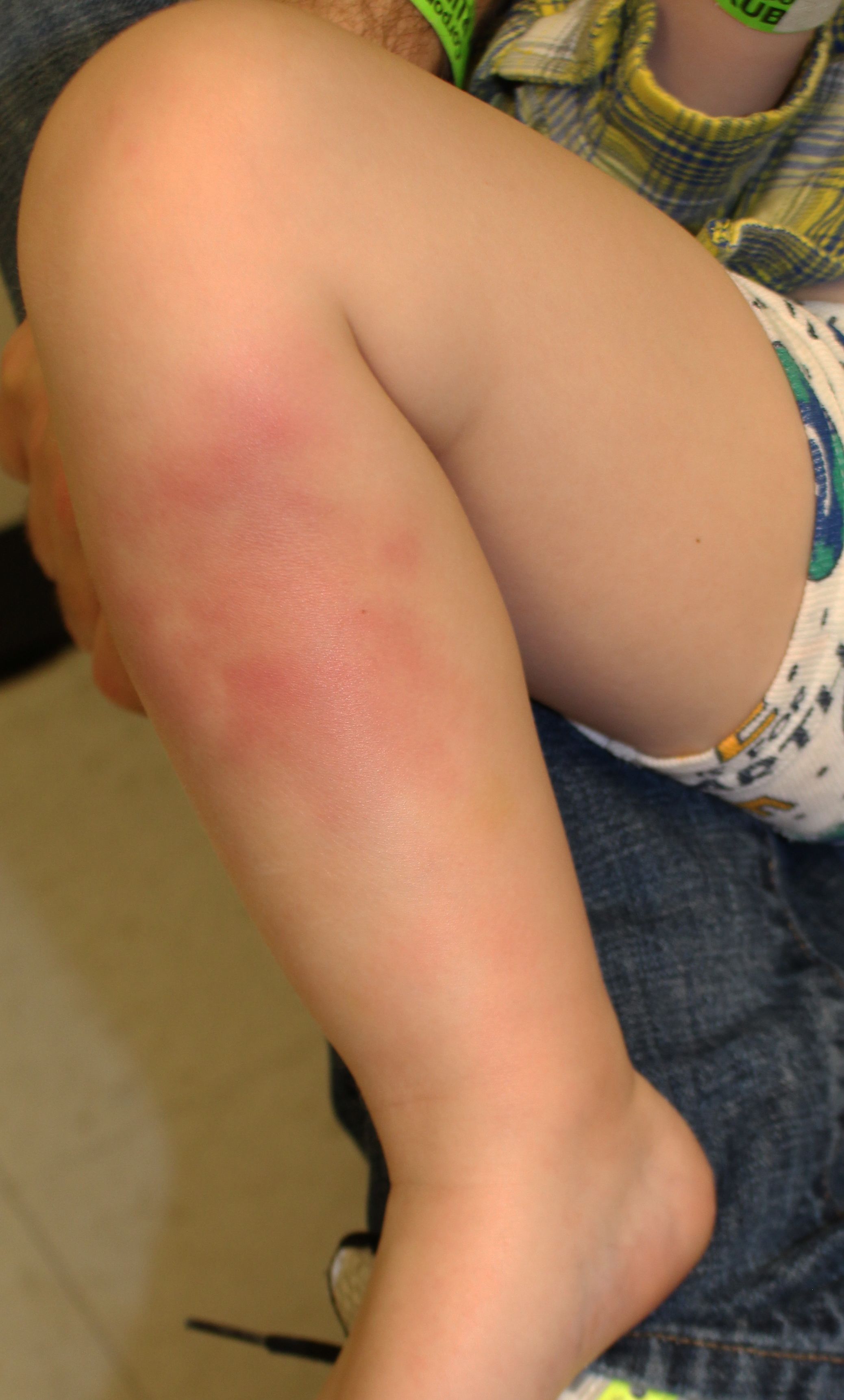
Painful red lumpy leg
A healthy 4-year-old boy presents with painful deep-seated bumps on the front of his right leg. He also complains of a sore throat for the last 4 days. What's the diagnosis?
The Case
You are asked to evaluate a healthy 4-year-old boy with painful deep-seated bumps on the front of his right leg. He did complain of a sore throat for the last 4 days.
Figure: Patient’s ventral right calf shows multiple erythematous subcutaneous nodules sensitive to touch

Diagnosis: Erythema Nodosum (EN) associated with strep throat
Discussion
EN is the most common form of panniculitis and is characterized by tender erythematous nodules or plaques located on the anterior and lateral surfaces of the lower legs.1,2 In children, EN is more commonly found on other parts of the body including the anterior thighs, upper limbs, trunk, and face.3 The full extent of lesions is usually better appreciated on palpation than visualized and most heal in 4-6 weeks without ulceration, scarring or suppuration.2,4 As lesions age, associated nodules soften and become less palpable.2
In children, EN is rare before 2 years old and peaks in teenagers.3 Although adult EN patients often present with symptoms of arthralgias and fever, arthralgias are less common in children.3 About half of all pediatric cases of EN are idiopathic.5 Of the remaining cases, EN is most commonly caused by Streptococcal and Yersinia infections.5
Adverse drug reactions to oral contraceptives and antibiotics have also been associated with EN. In children, hypersensitivity reactions to penicillin, sulfonamides, and macrolides have been linked to EN. Treatment involves discontinuation of the causative medication.6 In adolescent girls, oral contraceptives have been known to cause EN, reflecting a well-established association among EN, pregnancy, and hormonal therapy that has been known for decades.6 Diagnosis of EN due to contraceptives is made with disappearance of lesions concurrent with drug cessation.6 Other causes of EN include sarcoidosis and inflammatory bowel disease.7 However, these causes are much less common than infections especially in children.
Although the mechanism of EN manifestation is still unclear, it has been hypothesized to be due to a delayed type IV hypersensitivity reaction in subcutaneous fat. Subsequent inflammation of subcutaneous fat is facilitated by immune complex deposition in subcutaneous fat nodules and granuloma formation.5
Differential diagnosis
More specifically, EN is a septal panniculitis that should be distinguished from other diseases presenting with EN-like lesions such as Behcet's disease, nodular vasculitis, polyarteritis nodosa, erythema induratum and infectious cellulitis.2,3,4 Although not usually necessary in otherwise healthy children where the cause is often identified and the course is self-limited, histopathology of an incisional biopsy extending to the level above the fascia is recommended for a specific diagnosis.8 Early findings of EN will present with neutrophilic and eosinophilic inflammation mainly in and around septa usually without vasculitis. Later findings of EN will demonstrate a shift towards a lymphohistiocytic infiltrate.8 In contrast, EN-like lesions of Behcet's disease and nodular vasculitis will demonstrate lobular inflammation and lymphocytic or neutrophilic vasculitis.4
Erythema induratum presents with EN-like nodules and plaques but classically on the posterior aspect of the lower legs on the calves. Unlike EN, it is a lobar panniculitis presenting with vasculitis that can be distinguished by histopathology.2 In addition, erythema induratum is often associated with an underlying Mycobacterium tuberculosis infection, demonstrated by adjacent granulomatous panniculitis. Staining of Erythema induratum lesions may show acid-fast bacilli unlike in EN.2
Management of EN
While most cases of EN are self-limiting, treatment may be necessary for discomfort and/or underlying diseases. Leg elevation, rest, compression, and acetaminophen can be effective for discomfort. In cases of streptococcal or yersinia infections, antibiotic therapy may be necessary.5,8
Other symptomatic treatments include analgesics such as paracetamol and acetylsalicylic acid. For very painful lesions, nonsteroidal anti-inflammatory drugs such as naproxen and ketoprofen may be more effective.1
Patient outcome
A throat culture was positive for Group A beta-hemolytic strep, he was treated with oral amoxicillin for 10 days, and the skin lesions cleared within a week.
References
1. Kakourou T, Drosatou P, Psychou K, Aroni K, Nicolaidou P. Erythema nodosum in children: a prospective study. J Am Acad Dermatol. 2001 Jan;44(1):17-21 doi:10.1067/mjd.2001.110877.
2. Malaviya A, Abdeend S, Qurtom M, Al-Ghurier S, Kablawi H. Erythema nodosum or erythema induratum: an important distinction for appropriate treatment. Med Principles Pract. 1998;7:298–305 doi:10.1159/000026058
3. Labbé L, Perel Y, Maleville J, Taïeb A. “Erythema nodosum in children: a study of 27 patients. Pediatr Dermatol. Nov-Dec 1996;13(6):447-50. doi: 10.1111/j.1525-1470.1996.tb00722.x.
4. Yi S, Kim E, Kang H, Kim Y, Lee E. Erythema nodosum: clinicopathologic correlations and their use in differential diagnosis.” Yonsei Med J. 2007 Aug 31; 48(4): 601–608 doi: 10.3349/ymj.2007.48.4.601
5. Hafsi W, Badri T. Erythema nodosum. Updated December 3, 2021. Accessed February 23, 2022. www.ncbi.nlm.nih.gov/books/NBK470369/.
6. Schwartz R, Nervi S. Erythema nodosum: a sign of systemic disease. Am Fam Physician. 2007 Mar 1;75(5):695-700.
7. Starba A, Chowaniec M, Wiland P. Erythema nodosum - presentation of Three Cases.” Reumatologia. 2016; 54(2): 83–85. doi: 10.5114/reum.2016/60218
8. Blake T, Manahan M, Rodins K. Erythema nodosum – a Review of an uncommon panniculitis. Dermatol Online J. 2014 16;20(4):22376.
Recognize & Refer: Hemangiomas in pediatrics
July 17th 2019Contemporary Pediatrics sits down exclusively with Sheila Fallon Friedlander, MD, a professor dermatology and pediatrics, to discuss the one key condition for which she believes community pediatricians should be especially aware-hemangiomas.