Persistent birthmark grows on a toddler’s back
The parents of a healthy 20-month-old boy ask for advice about a birthmark on his lower back. The lesion is asymptomatic and has grown proportionately with their son. What's the diagnosis?
Figure 1

Figure 2

The case
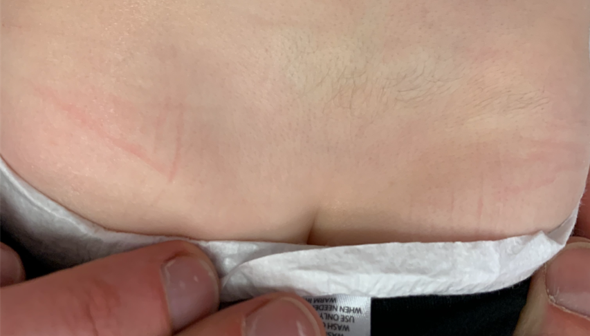
The parents of a healthy 20-month-old boy ask for advice about a birthmark on his lower back (Figure 1). The lesion is asymptomatic and has grown proportionately with their son.
Diagnosis: Congenital smooth muscle hamartoma
Clinical findings
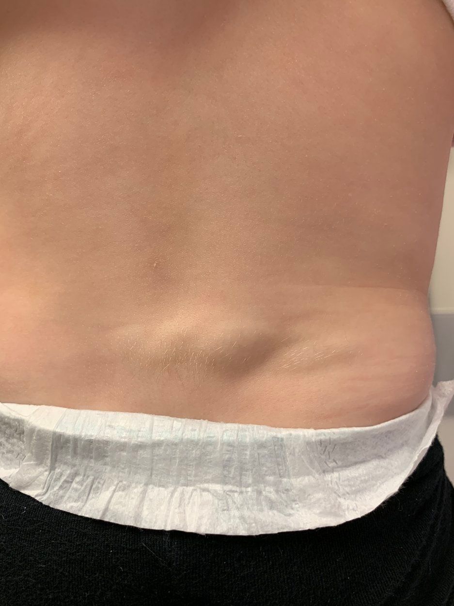
The child is noted to have a soft, partially compressible, nontender, skin-colored smooth plaque measuring approximately 5.5 cm by 3 cm with overlying hypertrichosis extending from the midline lower lumbar back to the middle-right lower lumbar back. Upon stroking the lesion, it becomes more raised and indurated transiently (Figure 2). There are no other similar growths anywhere else on the body. An ultrasound performed at an outside hospital revealed no underlying spinal abnormalities.
Etiology/Epidemiology
Smooth muscle hamartoma (SMH) is an innocent cutaneous lesion that can be categorized into congenital or acquired types.1 The congenital form of SMH is generally sporadic, although a familial case has been described.2,3 Smooth muscle hamartoma is occasionally referred to as “arrector pili hamartoma” because the malformation arises from disorganized proliferation of the smooth muscle cells in the arrector pili muscle.1,3,4 The etiology of SMH is unclear, but it likely arises secondary to a developmental anomaly of that tissue.5
The involvement of the arrector pili muscle is responsible for the characteristic “pseudo-Darier sign” in which the lesion becomes indurated or elevated upon stimulation of that muscle layer through stroking of the lesion.1,3-6 The patient may report a similar presentation when exposed to cold.3,5 The presence of this sign is suggestive of retained functional activity of the smooth muscle fibers and can distinguish SMH from similarly appearing lesions such as a congenital melanocytic nevus, potentially obviating the need for removal of this benign malformation.3 On histology, SMH appears as disseminated proliferation of smooth muscle cells in large, randomly oriented bundles usually located in the reticular dermis.3,5
The prevalence of SMH has been estimated at between 1 in 1000 and 1 in 2700 live births, with a reported male predominance in its development.3,5 It often presents with hypertrichosis and/or hyperpigmentation, although these features tend to diminish with age.3-6
Smooth muscle hamartoma usually presents as a solitary lesion, although it has occasionally been reported in a diffuse pattern associated with other complications as part of the Michelin tire baby syndrome (MTBS).5
Differential diagnosis
Smooth muscle hamartoma can be confused with congenital melanocytic nevus, which also can present as a raised lesion with hyperpigmentation and hypertrichosis, especially when the pseudo-Darier sign is absent.3 On dermoscopy, the patterns found in pigmented nevi are not present. Biopsy of the lesion should be performed prior to excision because selective congenital melanocytic nevi may need to be removed for their malignant potential whereas SMH is fully benign,3,5 and SMH is easily distinguishable from congenital melanocytic nevus on histopathology.3
Smooth muscle hamartoma also can masquerade as Becker melanosis and have a similar clinical and histologic appearance to those lesions.1,3 Although hypertrichosis, hyperpigmentation, and smooth muscle proliferation are seen in both lesions, smooth muscle proliferation is dominant in SMH whereas Becker melanosis has more robust hypertrichosis and hyperpigmentation, which both tend to worsen with age unlike in SMH.1,3 The 2 lesions can be differentiated by long-term follow-up to see whether worsening or improvement in the hypertrichosis and hyperpigmentation occurs.1
Treatment and management
Patients with clinically ambiguous lesions should have an evaluation by a dermatologist, and some lesions may require a biopsy to make a specific diagnosis.1,3-6 Parents should be reassured that the SMH lesion is benign and does not require treatment. They also should be advised that any hypertrichosis or hyperpigmentation will improve over time.3,5 If cosmetic therapy is desired by the parents or patient, treatment can include laser therapy to reduce the burden of hypertrichosis and hyperpigmentation or surgical excision.3
Patient outcome
The patient’s parents were reassured that the finding was a common benign lesion and had no malignant potential. They were advised that the lesion would likely continue to grow proportionately with the child, that it could be removed if it became bothersome, and that routine monitoring would be sufficient for now.
References:
1. Slifman NR. Harrist TJ, Rhodes AR. Congenital arrector pili hamartoma. A case report and review of the spectrum of Becker's melanosis and pilar smooth-muscle hamartoma. Arch Dermatol. 1985;121(8):1034-1037.
2. Gualandri L, Cambiaghi S, Ermacora E, Tadini G, Gianotti R, Caputo R. Multiple familial smooth muscle hamartomas. Pediatr Dermatol. 2001;18(1):17-20.
3. Schmidt CS, Bentz ML. Congenital smooth muscle hamartoma: the importance of differentiation from melanocytic nevi. J Craniofac Surg. 2005;16(5):926-929.
4. Bilgiç Ã, Tunçez Akyürek F, Altinyazar HC. Pseudo Darier sign: a distinctive finding for congenital smooth muscle hamartoma. J Pediatr. 2016;169:318.
5. Raboudi A, Litaiem N. Congenital smooth muscle hamartoma. StatPearls [Internet]. Updated August 11, 2019. Accessed May 1, 2020. https://www.ncbi.nlm.nih.gov/books/NBK545188/
6. Goldman MP, Kaplan RP, Heng MC. Congenital smooth-muscle hamartoma. Int J Dermatol. 1987;26(7):448-452.
Recognize & Refer: Hemangiomas in pediatrics
July 17th 2019Contemporary Pediatrics sits down exclusively with Sheila Fallon Friedlander, MD, a professor dermatology and pediatrics, to discuss the one key condition for which she believes community pediatricians should be especially aware-hemangiomas.