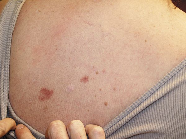
Persistent pearly plaques in a fair-skinned adolescent girl

FIGURE Two pearly round red plaques on an adolescent girl.
The Case
You are asked to evaluate 2 persistent, pearly red plaques on the back of a healthy 19-year-old girl. Her mother says she saw them last week when her children were walking to the neighborhood pool. The patient says that the plaques have been present for at least the last 6 months but have not bothered her. Of note, she is active in outdoor sports and works as a lifeguard and swimming instructor on weekends during the summer. What’s the diagnosis?
Diagnosis:
Basal cell carcinoma (BCC)
Epidemiology
Basal cell carcinoma is a common skin cancer arising from the basal layer of the epidermis. These cancers have low metastatic potential but may become locally invasive and disfiguring. The American Cancer Society estimates that more than 2 million nonmelanoma skin cancers were treated in the United States in 2006, of which the majority were BCCs.1 The incidence of BCC has increased by more than 10% per year, particularly among American women aged younger than 40 years.2
Risk factors
Exposure to natural and artificial ultraviolet radiation is the most important risk factor. Patient-specific risk factors include fair skin, light-colored eyes, red hair, childhood freckling, use of tanning beds, and increased number of past sunburns.3,4 Although basal cell carcinomas may occur in the pediatric population in the context of cancer-related genodermatoses such as basal cell nevus syndrome, they can also occur independently in high-risk patients with light skin and increased sun exposure.
Molecular pathogenesis
Basal cell carcinoma is thought to derive from the basaloid epithelia where pluripotent progenitor epithelia arise. Mutations in PTCH1 and BCL2 have been implicated in tumorigenesis.5
Typical BCCs are pearly pink or flesh-colored papules and plaques with overlying telangiectasia. Lesions may be translucent or slightly erythematous with a violaceous or pearly rolled border, sometimes accompanied by scaling, crusting, and/or ulceration. Approximately 80% of BCCs occur on the head and neck with the remainder occurring mainly on the trunk and lower limbs.6
Differential diagnosis
Early nodular variants of BCC may look like benign growths such as dermal nevi, small epidermal inclusion cysts, or sebaceous hyperplasia. Lesions may also resemble Molluscum contagiosum and amelanotic melanoma. Larger lesions may resemble squamous cell carcinoma and keratoacanthomas.
Diagnosis and treatment
The diagnosis can often be made upon the clinical examination with skin biopsy for histological confirmation. Tumor characteristics including size, location, and pathology guide treatment. Basal cell carcinomas at low risk for recurrence are most commonly managed with electrodesiccation and curettage or surgical excision. Topical 5-fluorouracil, imiquimod, cryosurgery, and photodynamic therapy may also be employed, although less frequently. Patient-specific factors related to the ability to tolerate surgery and preference should also play a role.
Prognosis
The prognosis is excellent for most patients with BCCs, which are slow growing and rarely metastasize. A third of patients with 1 BCC develop another primary lesion within 1 year. There is increased risk for development of subsequent squamous cell carcinoma and melanoma in patients with a history of BCC, necessitating close follow-up care.7 Early detection may mean detecting smaller, less-aggressive tumors with good treatment outcomes. Patients should also be educated on sun avoidance, use of broad-spectrum sunscreen (UVA and UVB), wearing sun-protective clothing, avoiding tanning salons, and performing monthly skin self-exams, by the patients themselves or by a family member.
REFERENCES
1. American Cancer Society. Cancer Facts and Figures 2010. Atlanta, GA: American Cancer Society; 2010. http://www.cancer.org/acs/groups/content/@epidemiologysurveilance/documents/document/acspc-026238.pdf. Accessed June 5, 2013.
2. Christenson LJ, Borrowman TA, Vachon CM, et al. Incidence of basal cell and squamous cell carcinomas in a population younger than 40 years. JAMA. 2005;294(6):681-690.
3. Ferrucci LM, Cartmel B, Molinaro AM, Leffell DJ, Bale AE, Mayne ST. Indoor tanning and risk of early-onset basal cell carcinoma. J Am Acad Dermatol. 2012;67(4):552-562.
4. van Dam RM, Huang Z, Rimm EB, et al. Risk factors for basal cell carcinoma of the skin in men: results from the health professionals follow-up study. Am J Epidemiol. 1999;150(5):459-468.
5. Fan H, Oro AE, Scott MP, Khavari PA. Induction of basal cell carcinoma features in transgenic human skin expressing Sonic Hedgehog. Nat Med. 1997;3(7):788-792.
6. Wong CS, Strange RC, Lear JT. Basal cell carcinoma. BMJ. 2003;327(7418):794-798.
7. Schreiber MM, Moon TE, Fox SH, Davidson J. The risk of developing subsequent nonmelanoma skin cancers. J Am Acad Dermatol. 1990;23(6 pt 1):1114-1118.
MR CHEN is a third-year medical student at Johns Hopkins University School of Medicine, Baltimore, Maryland. DR COHEN, the section editor for Dermatology: What’s Your Dx?, is director, Pediatric Dermatology and Cutaneous Laser Center, and associate professor of pediatrics and dermatology, Johns Hopkins University School of Medicine, Baltimore. The author and section editor have nothing to disclose regarding affiliations with or financial interests in any organizations that may have an interest in any part of this article. Vignettes are based on real cases that have been modified to allow the author and editor to focus on key teaching points. Images may also be edited or substituted for teaching purposes.
Recognize & Refer: Hemangiomas in pediatrics
July 17th 2019Contemporary Pediatrics sits down exclusively with Sheila Fallon Friedlander, MD, a professor dermatology and pediatrics, to discuss the one key condition for which she believes community pediatricians should be especially aware-hemangiomas.