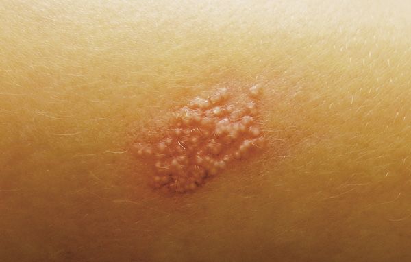
Posttraumatic lesion persists on child’s forearm
A worried mother brings her 2-year-old boy to your office for evaluation of an asymptomatic skin eruption that has been present for 2 months. The lesion developed 6 months after he sustained an abrasion to the same site when he fell on concrete steps.
The Case
A worried mother brings her 2-year-old boy to your office for evaluation of an asymptomatic skin eruption that has been present for 2 months. The lesion developed 6 months after he sustained an abrasion to the same site when he fell on concrete steps.

The patient's right forearm displays 1.5 cm x 0.8 cm erythematous plaque studded with firm white 1 mm-to-2 mm papules.
DERMCASE diagnosis
Milia en plaque
Discussion
Milia en plaque is a rare but benign dermatologic condition characterized by grouped milia on an erythematous base.1 It typically affects middle-aged women and presents with a periauricular distribution.2 Clinically the lesions are asymptomatic, arise spontaneously, and may regress without treatment. Diagnosis is based on recognition of clinical findings and biopsy can be confirmatory but is rarely required. The pathogenesis remains unknown.
Only 9 cases of milia en plaque have been reported in the child and adolescent population.3-9 Those lesions arose spontaneously and their distributions were confined to facial areas, most commonly the periorbital region. This is the first case of extrafacial milia en plaque to be reported in the pediatric literature and, additionally, the youngest patient to our knowledge to be diagnosed. This case is also unique because the milia en plaque developed at the site of previous trauma, suggesting that secondary milia en plaque can also occur.
It is well recognized that isolated milia can arise as a part of scar formation,1 suggesting that this case is likely a rare variant of a common posttraumatic reaction. Given the location of the lesion and its posttraumatic occurrence, it is reasonable to consider diagnoses such as a secondary bacterial or viral infection in this patient. However, the long-standing firm papules and lack of associated symptoms are not characteristic of a secondary infection.
Treatment
The child in this presentation was initially treated with an oral antibiotic as well as topical mupirocin by his pediatrician without any improvement. It is therefore important to be able to recognize milia en plaque and consider it as part of a differential diagnosis because management differs considerably.
The most commonly used conservative option in the pediatric population is use of a topical retinoid such as tretinoin cream, or observation only, given that this is a harmless, asymptomatic entity. One previous case reported a response to oral minocycline treatment after topical medications had been trialed.5 If the lesions persist despite conservative treatment or become bothersome, or if the lesions are in a cosmetically undesirable location, simple extraction can also be considered in the appropriate patient.
Our patient
The boy in this case was provided with a prescription for tretinoin 0.1% cream to apply 3 nights per week, and was given the option of future extraction if the milia persist.
Conclusion
Milia en plaque is an uncommon dermatologic condition that has been described infrequently, with even fewer cases noted in the pediatric population. It is important to recognize this entity, arising either as a primary lesion or secondary to an insult such as trauma, for proper diagnosis and treatment. This case demonstrates that milia en plaque can occur in patients aged as young as 21 months and involve sites of the body other than the face.
REFERENCES
1. Berk DR, Bayliss SJ. Milia: a review and classification. J Am Acad Dermatol. 2008;59(6):1050-1063.
2. Stefanidou MP, Panayotides JG, Tosca AD. Milia en plaque: a case report and review of the literature. Dermatol Surg. 2002;28(3):291-295.
3. Lee DW, Choi SW, Cho BK. Milia en plaque. J Am Acad Dermatol.1994;31(1):107.
4. Bouassida S, Meziou TJ, Mlik H, et al. Childhood plaque milia of the inner canthus. Ann Dermatol Venereol.1998;125(12):906-908.
5. Bridges AG, Lucky AW, Haney G, Mutasim DF. Milia en plaque of the eyelids in childhood: case report and review of the literature. Pediatr Dermatol. 1998;15(4):282-284.
6. Dogra S, Kanwar AJ. Milia en plaque. J Eur Acad Dermatol Venereol. 2005;19(2):263-264.
7. Cota C, Sinagra J, Donati P, Amantea A. Milia en plaque: three new pediatric cases. Pediatr Dermatol. 2009;26(6):717-720.
8. Boggio P, Alperovich R, Spiner RE, Schroh R, Hassan M. Letter: Periorbital bilateral milia en plaque in a female teenager. Dermatol Online J. 2012;18(4):11.
9. Zhang RZ, Zhu WY. Bilateral milia en plaque in a 6-year-old Chinese boy. Pediatr Dermatol. 2012;29(4):504-506.
Dr Thorn is PGY-1, Department of Pediatrics, Johns Hopkins University School of Medicine, Baltimore, Maryland. Dr Kirkorian is instructor of dermatology, Department of Dermatology, Johns Hopkins University School of Medicine, Baltimore. Dr Grossberg is assistant professor of dermatology, Department of Dermatology, Johns Hopkins University School of Medicine, Baltimore. Dr Cohen, section editor for Dermcase, is professor of pediatrics and dermatology, Johns Hopkins University School of Medicine, Baltimore. The authors and section editor have nothing to disclose in regard to affiliations with or financial interests in any organizations that may have an interest in any part of this article. Vignettes are based on real cases that have been modified to allow the authors and section editor to focus on key teaching points. Images also may be edited or substituted for teaching purposes.
Recognize & Refer: Hemangiomas in pediatrics
July 17th 2019Contemporary Pediatrics sits down exclusively with Sheila Fallon Friedlander, MD, a professor dermatology and pediatrics, to discuss the one key condition for which she believes community pediatricians should be especially aware-hemangiomas.