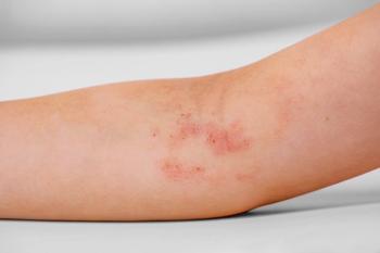
Purple papules pop up after antibiotic course
The mother of a 17-year-old boy frantically brings him to the office for evaluation of an itchy, burning rash that started on his hands 1 hour ago.
Annular lesions with well-demarcated, dusky, violaceous plaques with peripheral erythema erupted on the hands of a 17-year-old boy.
THE CASE
The mother of a 17-year-old boy frantically brings him to the office for evaluation of an itchy, burning rash that started on his hands 1 hour ago. Last night, her son started a course of trimethoprim-sulfamethoxazole for traveler’s diarrhea. She says that he had some of the exact same purple spots in the same location last year when he was given the same medication for a similar complaint, but there are also new lesions this time. Read more >>>
DERMCASE DIAGNOSIS Fixed drug eruption
Fixed drug eruptions (FDEs) are common among cutaneous drug eruptions with a typical history of recurrent lesions in the same location upon repeated exposure to the offending drug.1 The clinical findings are classically described as annular or target lesions with well-demarcated, dusky, violaceous plaques with peripheral erythema and sometimes central bulla formation. More than 700 drugs are known to cause FDEs with the most common agent reported being trimethoprim-sulfamethoxazole.2
Pathophysiology and etiology
In addition to trimethoprim-sulfamethoxazole, tetracyclines and nonsteroidal anti-inflammatory agents are the most common triggers.3 It is thought that FDEs are caused by a type IV cell-mediated hypersensitivity reaction. When the body is exposed to the offending drug, CD8-T cells are stimulated to produce interferon gamma, a cytokine that initiates an inflammatory response. The CD4-T cells as well as neutrophils are recruited to the site and contribute to tissue damage and development of the characteristic cutaneous lesions. The T-cells are fixed to the site of involvement resulting in the same area of involvement on reexposure to the inciting medication.
Familial cases have been reported and previous studies suggest a possible genetic predisposition to FDEs related to HLA-B22.4 A specific link between HLA-A30 B13 Cw6 haplotype and trimethoprim-sulfamethoxazole-induced FDEs also has been reported.5
Epidemiology
Fixed drug eruptions are the second most common cause of cutaneous drug eruptions in children and adults after exanthematous drug eruptions.1 Previously reported studies have estimated the prevalence of FDEs in children as between 20% to 50% of cutaneous drug eruptions.
Clinical presentation
Within 30 minutes to 8 hours postingestion of the offending drug, patients can develop annular erythematous to violaceous plaques, some with classic target lesions.3 Plaques most commonly develop on the lips, hands, oral mucosa, and genitalia.1 The lesions can be single or multiple and progress from a well-defined erythematous stage to a hyperpigmented stage that can last for weeks to months.3
As lesions begin to heal, crusting and scaling can occur along with a dusky brown color at the site. Nonpigmented FDEs with exposure to pseudoephedrine hydrochloride and tetrahydrozoline have been described as lesions that fade without pigmentation.6
Differential diagnosis
Fixed drug eruptions are more common in adults than children.3 Differential diagnosis includes insect bites, urticaria, erythema multiforme, erythema dyschromicum perstans, and erythema annulare centrifugum.1 Severe FDEs can have associated blistering and can be misdiagnosed as autoimmune or mechanical blistering disorders.1,2
The diagnosis of FDE is made based on a good history of development of lesions in relation to taking the offending drug. Although the history and clinical findings are usually diagnostic, if the diagnosis is unclear, skin biopsy may be done, which will show a lichenoid infiltrate, hydropic degeneration of the basal cell layers, and dyskeratotic keratinocytes.2 During the pigmented stage of lesions, biopsy may show melanin-filled macrophages.
Immunohistochemical studies may show CD8 and CD3 staining for T cells along the basal layer and upper dermis with a mixed inflammatory infiltrate.3 Patch testing also has been reported to aid in identifying a causative agent by applying the most common offending drugs to the child’s upper back using Finn Chambers and occluding for 48 hours. Systemic provocation also can be done, but because of the risk of severe and generalized reactions, patch testing may be a safer alternative.
Treatment and medications
The best and most simple treatment is to identify and remove the offending drug.3 Aside from this, treatment is largely symptomatic. Systemic antihistamines and topical corticosteroids can be used if needed.
Patient outcome
The patient developed characteristic annular, erythematous to violaceous target-shaped lesions on his fingers every time he took trimethoprim-sulfamethoxazole. Given the patient’s history of recurrent lesions in the exact same spot after exposure to the offending drug, an FDE was considered most likely, and the patient was advised to avoid taking this antibiotic in the future. His lesions eventually resolved, leaving behind residual hyperpigmentation that will probably fade somewhat within weeks to months.
REFERENCES
1. Can C, Akkelle E, Bay B, Arican O, Yalçin O, Yazicioglu M. Generalized fixed drug eruption in a child due to trimethoprim/sulfamethoxazole. Pediatr Allergy Immunol, 2014;25(4):413-415.
2. Morelli JG, Tay YK, Rogers M, Halbert A, Krafchik B, Weston WL. Fixed drug eruptions in children. J Pediatr, 1999;134(3):365-367.
3. Ozkaya-Bayazit E, Akar U. Fixed drug eruption induced by trimethoprim-sulfamethoxazole: evidence for a link to HLA-A30 B13 Cw6 haplotype. J Am Acad Dermatol. 2001;45(5)712-717.
4. Pellicano R, Ciavarella G, Lomuto M, Di Giorgio G. Genetic susceptibility to fixed drug eruption: evidence for a link with HLA-B22. J Am Acad Dermatol. 1994;30(1):52-54.
5. Shelley WB, Shelley ED. Nonpigmenting fixed drug eruption as a distinctive reaction pattern: examples caused by sensitivity to pseudoephedrine hydrochloride and tetrahydrozoline. J Amer Acad Dermatol. 1987;17(3)403-407.
6. Shin, HT, Chang MW. Drug eruptions in children. Curr Probl Pediatr. 2001;31(7):207-234.
Mr Singla is a fourth-year medical student at the Brody School of Medicine at East Carolina University, Greenville, North Carolina. Dr Cohen, section editor for Dermcase, is professor of pediatrics and dermatology, Johns Hopkins University School of Medicine, Baltimore, Maryland. The author and section editor have nothing to disclose in regard to affiliations with or financial interests in any organizations that may have an interest in any part of this article. Vignettes are based on real cases that have been modified to allow the author and section editor to focus on key teaching points. Images also may be edited or substituted for teaching purposes.
Newsletter
Access practical, evidence-based guidance to support better care for our youngest patients. Join our email list for the latest clinical updates.


