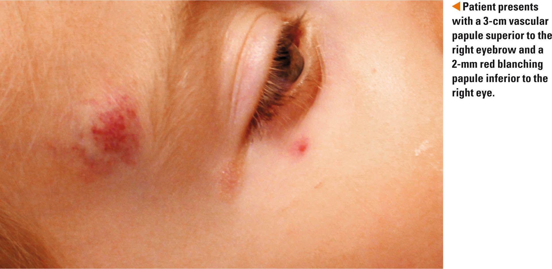
Is this red papule another hemangioma?
The parents of a 3-year-old girl with a history of a slowly regressing infantile hemangioma on her right forehead were afraid that she was developing a new hemangioma near her right eye.

THE CASE
The parents of a 3-year-old girl with a history of a slowly regressing infantile hemangioma on her right forehead were afraid that she was developing a new hemangioma near her right eye. The blanching red papule had been slowly increasing in size for 2 months. CLICK FOR MORE ON THIS CASE.
DERMCASE diagnosis: Spider angioma
The differential diagnosis includes spider angioma, pyogenic granuloma, and infantile hemangioma.
Spider angioma
A spider angioma, or nevus araneus, typically presents with a 2-mm to 3-mm red macule or slightly elevated papule representing a central feeder artery with radiating dilated vessels (spider legs) and surrounding flushing.1 Blanching occurs when pressure is applied to the central artery and radiating vessels. Spider angiomas are generally found during times of elevated levels of estrogens including cirrhosis and pregnancy. They also develop in 15% of preschoolers and 45% of school-aged children and usually regress on their own following puberty. The most common sites include the hand, forearm, face, and ears. Although treatment is not necessary, pulse dye laser is the first-line therapy if the patient desires treatment or if the lesions are in areas prone to trauma.
More: A 12-year-old with a perplexing rash
Pyogenic granuloma
Pyogenic granulomas (PGs) typically develop as 2-mm to 5-mm red, glistening, sessile or pedunculated papules in infants after the neonatal period, and also in children and adults. They often have a discernible scaly, crusted border that helps to distinguish them from small hemangiomas. In many cases, PGs appear at sites of injury. They demonstrate rapid growth in the beginning and may ulcerate or bleed when traumatized. The areas most prone for development of PGs include the face, arms, and hands.
Treatment for small granulomas includes flashlamp-pumped pulse dye laser. Some granulomas may regress with silver nitrate cauterization.1 Larger lesions require excision and electrodessication of the base. More recent anecdotal reports suggest that uncomplicated lesions may resolve with topical beta-blocker liquid drops (timolol/Timoptic).2
Infantile hemangioma
Infantile hemangiomas (IHs) are the most common benign vascular tumors that arise in the neonatal period.3,4 They are typically divided into 3 subtypes: superficial, deep, and mixed.3 The superficial hemangiomas are located in the upper dermis and usually present as red macules, papules, and plaques. Deep hemangiomas are found in the deep dermis and subcutaneous fat, and mixed lesions demonstrate features of both deep and superficial lesions. Solitary round or oval lesions are referred to as focal IH, similar multiple lesions as multiple focal IH, and larger patterned lesions as segmental IH. There is a predominance in females, and also in Caucasian infants and in those born prematurely or weighing less than 1000g.4
Hemangiomas usually appear in the first few days to first few weeks of life and may grow for 6 to 12 months. Many peak at 3 to 4 months and begin to regress after 6 months.3 Clinically, infantile hemangiomas can present with precursor lesions such as telangiectasias with a surrounding halo, pale areas, pink macules, and bluish-colored bruise-like areas.4 Infantile hemangiomas can develop on any skin or mucosal surface, with 50% occurring on the head and neck. The most common complication that can occur in 10% of infantile hemangiomas is ulceration. Other complications include obstruction of vital structures, high-output cardiac failure, and disfigurement secondary to size and location.
Treatment options depend on the severity of the IH and location. Often, only close observation and education for the family is necessary. Ulcerated hemangiomas may require appropriate wound care. Topical beta-blockers have been used for small superficial IH, whereas oral beta-blockers have become the standard of care for complicated IH.
Our patient
The IH on our patient’s forehead was involuting, so we opted to monitor it for continued improvement. The spider angioma was not symptomatic. We discussed possible laser therapy, but the parents decided to have us monitor for now as well.
REFERENCES
1. Morelli JG. Vascular disorders. In: Kliegman RM, Stanton BF, St Geme JW III, Schor NF, Behrman RE. Nelson Textbook of Pediatrics. 19th ed. Philadelphia, PA: Elsevier Saunders; 2011:2223-2231.
2. Wine Lee L, Goff KL, Lam JM, et al. Treatment of pediatric pyogenic granulomas using β-adrenergic receptor antagonists. Pediatr Dermatol. 2014;31(2):203-207.
3. Wahrman JE, Honig PJ. Hemangiomas. Pediatr Rev. 1994;15(7);266-271.
4. Haggstrom AN, Garzon MC. Infantile hemangiomas. In: Bolognia JL, Jorizzo JL, Schaffer JV. Dermatology. 3rd ed. Philadelphia, PA: Elsevier Saunders; 2012:1691-1709.
Dr Richardson is a senior pediatric resident at the Herman and Walter Samuelson Children’s Hospital, Baltimore, Maryland. She has accepted a position as a primary pediatrician in Waldorf, Maryland. Dr Cohen, section editor for Dermcase, is professor of pediatrics and dermatology, Johns Hopkins University School of Medicine, Baltimore, Maryland. The author and section editor have nothing to disclose in regard to affiliations with or financial interests in any organizations that may have an interest in any part of this article. Vignettes are based on real cases that have been modified to allow the author and section editor to focus on key teaching points. Images also may be edited or substituted for teaching purposes.
Recognize & Refer: Hemangiomas in pediatrics
July 17th 2019Contemporary Pediatrics sits down exclusively with Sheila Fallon Friedlander, MD, a professor dermatology and pediatrics, to discuss the one key condition for which she believes community pediatricians should be especially aware-hemangiomas.