Rings around the nevi
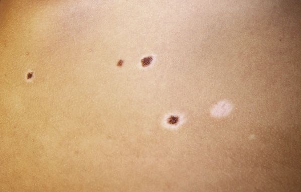
A panicked mother of an 11-year-old girl brings her daughter to your office for evaluation of changing moles that she noted when they returned from the family beach vacation last weekend. What's the diagnosis?

The Case
A panicked mother of an 11-year-old girl brings her daughter to your office for evaluation of changing moles that she noted when they returned from the family beach vacation last weekend. What’s the diagnosis?
Diagnosis:
Halo nevus
Clinical features
Halo nevus, also termed leukoderma acquisitum centrifugum or Sutton nevus, is a benign pigmented melanocytic nevus surrounded by a depigmented ring.1,2 Halo nevi have an incidence of 1% in the general population. The mean age of onset is 15 years, and they occur equally in males and females. The nevus itself is most commonly acquired, but rarely develops around congenital nevi. Patients may have 1 or multiple halo nevi, and the most common location is on the trunk.
Halo nevi have 4 clinical stages of development.1,2 In the first stage, a pigmented nevus is present surrounded by the characteristic halo of depigmentation. Stage 2 occurs when the nevus loses its pigmentation, leaving a skin-colored macule or papule surrounded by depigmentation. In stage 3, the papule or macule in the center vanishes completely, with only an oval or round patch of depigmentation remaining. In the final stage, repigmentation of the area occurs, resulting in the disappearance of the halo nevus completely. Each individual halo nevus may go through all 4 stages or may cease development at any stage. Nevi that do progress through the fourth stage may take years or even decades to completely repigment.1
Halo nevi may be seen more frequently in patients with Turner syndrome and vitiligo.2 About 20% of children and adults with vitiligo have halo nevi, and those with multiple halo nevi are more likely to have concurrent vitiligo. Although the causes of both vitiligo and halo nevi are poorly understood, both are most commonly believed to be an immune-mediated process resulting from damage or destruction of melanocytes.2,3 Family history may be positive for halo nevus, vitiligo, atopic dermatitis, and autoimmune disorders, most commonly Hashimoto thyroiditis.2
Etiology and pathology
While the precise etiology is unknown, autoimmune phenomenon is believed to play a role in the development of halo nevi.2,4 Skin biopsy shows a mononuclear infiltrate comprised primarily of macrophages, cytotoxic T cells, and Langerhans cells surrounding the nevus at the epidermal-dermal junction and within the papillary dermis.3 Histology of the halo of depigmentation demonstrates a decrease in the number of melanocytes and melanophages, as well as a decrease in pigment within keratinocytes.4
Differential diagnosis and treatment
The differential diagnosis for a halo nevus should include malignant melanoma, atypical nevus, and postinflammatory hypopigmentation.1,3 Although the risk of melanoma developing in conjunction with halo nevi is rare, the presence of a nevus with changing color or border requires careful evaluation. The central nevus should be examined closely, and a biopsy performed if warranted. Benign-appearing halo nevi warrant observation and periodic reexamination (eg, annually).
Our patient
The nevi in the centers of our patient’s halos appeared to be quite stable, and at a follow-up visit 6 months later they appeared unchanged. Photographs were obtained for continued monitoring, and her mother was reassured.
REFERENCES
1. Aouthmany M, Weinstein M, Zirwas MJ, Brodell RT. The natural history of halo nevi: A retrospective case series. J Am Acad Dermatol. 2012;67(4):582-586.
2. Patrizi A, Bentivogli M, Raone B, Dondi A, Tabanelli M, Neri I. Association of halo nevus/i and vitiligo in childhood: a retrospective observational study. J Eur Acad Dermatol Venereol. 2013;27(2):e148-e152.
3. Huynh PM, Lazova R, Bolognia J. Unusual halo nevi-darkening rather than lightening of the central nevus. Dermatology. 2001;202(4):324-327.
4. Tokura Y, Yamanaka K, Wakita H, et al. Halo congenital nevus undergoing spontaneous regression. Involvement of T-cell immunity in involution and presence of circulating anti-nevus cell IgM antibodies. Arch Dermatol. 1994;130(8):1036-1041.
IMAGE CREDIT/AUTHOR SUPPLIED
DR ETZLER is a resident at Albert Einstein Medical Center, Philadelphia, Pennsylvania. She will begin a 3-year residency in dermatology at Hahnemann University Hospital, Philadelphia, in 2014. DR COHEN, the section editor for Dermatology: What’s Your Dx?, is director, Pediatric Dermatology and Cutaneous Laser Center, and associate professor of pediatrics and dermatology, Johns Hopkins University School of Medicine, Baltimore, Maryland. The author and section editor have nothing to disclose regarding affiliations with or financial interests in any organization that may have an interest in any part of this article. Vignettes are based on real cases that have been modified to allow the author and editor to focus on key teaching points. Images also may be edited or substituted for teaching purposes.
Subscribe to Contemporary Pediatrics to get monthly clinical advice for today's pediatrician.