A scaly rash: A diagnosis that rings deep
A 6-year-old patient presents with an annular, scaly lesion on his leg. What could be the cause?
"All images courtesy of http://www.dermatlas.org/. Vignettes are based on real cases which have been modified to allow the authors and editor to focus on key teaching points. Images may also be edited or substituted for teaching purposes."

>> What's the diagnosis?
Clinical findings

Topical steroids and tinea infections
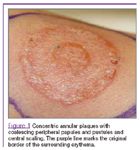
Application of topical steroids to unrecognized superficial fungal skin infections can have two important clinical consequences. First, it can mask the characteristic scaly, annular appearance of tinea corporis, and may lead to concentric expanding plaques, as seen in Figure 1. This altered morphology, known as tinea incognito, can make the diagnosis more challenging. Second, steroids can modify a patient's local immunologic response. This allows spreading into deeper tissues, and can lead to a dermatophytic folliculitis known as Majocchi's granuloma, as occurred in this patient.1-4
Etiology
Majocchi's granuloma was first described by Domenico Majocchi in 1883.5 Although the exact incidence is not known, it is well described in two populations. Immunocompromised patients, including those taking chronic steroids or with HIV,6,7 have an increased incidence. In otherwise healthy patients, Majocchi's granuloma results from opportunistic dermatophytic infection, usually in conjunction with the application of topical steroids2,3 or disruption of the skin barrier. This may come about through routine bumps and bruises in the pediatric population, or secondary to shaving in women.1,8
Majocchi's granuloma is generally preceded by a superficial fungal infection, which may involve the skin (tinea corporis), scalp (tinea capitis or kerion), feet (tinea pedis), or nails (onychomychosis). 1,4,5,9 Systemic immunosuppression or topical steroids as mild as 1% hydrocortisone interfere with antifungal activity and local cell-mediated immunity, allowing the fungi to invade deeper tissues.3,10 Invasion of the hair follicle or adjacent dermal tissue leads to a local suppurative folliculitis or perifolliculitis, and may progress to a granulomatous reaction.11
Recognize & Refer: Hemangiomas in pediatrics
July 17th 2019Contemporary Pediatrics sits down exclusively with Sheila Fallon Friedlander, MD, a professor dermatology and pediatrics, to discuss the one key condition for which she believes community pediatricians should be especially aware-hemangiomas.