Teen distressed by scaly papules and plaques
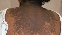
An obese 16-year-old girl has been depressed about a slowly progressive skin eruption on her chest and back for a year.
WHAT'S YOUR DX?
The Case

CLINICAL FINDINGS AND ETIOLOGY
Confluent and reticulated papillomatosis (CARP) was originally described by Gougerot and Carteaud in 1927.1 Although it is often described as a rare dermatosis, our experience suggests that it is much more common than reported. CARP can affect persons up to 40 years old but usually begins during puberty. It can occur in all ethnicities but tends to be more common in black patients and in females.2-4

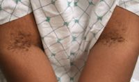
CARP often coexists with the velvety, dirty-brown plaques of acanthosis nigricans (AN), which is most prominent in the intertriginous folds and over bony prominences. Some authors have questioned whether CARP and acanthosis nigricans are indeed separate entities because of their clinical and histologic similarities.5 The etiology and pathogenesis of CARP are unclear. Proposed triggers include an endocrine disturbance, abnormal keratinization, an amyloidosis variant, a hereditary condition, or a host response to bacterial or fungal infection.3
DIFFERENTIAL DIAGNOSIS
AN presents as velvety, dark thickening of the skin marked by epidermal keratinocyte and dermal fibroblast proliferation and papillomatosis on histology. It is occasionally pruritic but usually asymptomatic. This condition can occur at any age, but onset is most frequent in teenagers and young adults. It is associated with increased insulin or insulin-like growth factor levels. In addition to occurring during puberty, when trophic hormone levels are high, it can be seen commonly in obese patients or in those with diabetes and insulin resistance. Although it can occur in any skin type, lesions are clinically more obvious in dark-pigmented persons. Although the eruption is generalized, the most prominent areas of involvement include the intertriginous areas of the axillae, groin, and neck. Mucous membranes are also affected, and skin tags in the intertriginous regions often accompany AN. Patients with AN should be evaluated for obesity and hyperinsulinemia.
Pseudoacanthosis nigricans, often confused with AN, is characterized by postinflammatory hyperpigmentation and thickened skin secondary to maceration of the skin caused by rubbing and increased sweating.
Tinea versicolor (TV) may also be confused with CARP. Clinically, TV consists of discrete and confluent 3- to 4-mm hypopigmented or hyperpigmented macules, with fine overlying scale. In many patients, the scaling can only be appreciated when the lesions are scraped with the examining fingernail. The eruption most commonly appears on the chest, back, abdomen, and proximal extremities, but it can also be distributed in the flexor creases and face. Some individuals develop red-brown papules with or without scale that may coalesce.
TV is associated with Malassezia species, which can be seen on skin scraping as spores and hyphae, and it often is brought to the attention of the physician when affected areas fail to tan during the summer. In temperate climates, TV is seen more commonly in the summer months when it is warmer and humid. In tropical countries, it is diagnosed year-round and in patients of all ages. However, in temperate climates, TV, like CARP, occurs most frequently in teenagers and young adults.
Many patients with CARP are misdiagnosed with TV, and the proper diagnosis is only considered when treatment for TV is unsuccessful. Although topical and oral antifungal medications may clear TV, CARP is resistant to these therapies.
Macular amyloidosis presents with variably pruritic, gray-brown macular or minimally elevated symmetrical eruption over the central back or arms. Lesions may be discrete or confluent. Although the cause is unknown, macular amyloidosis probably results from chronic injury to the skin in the setting of chronic itching. The disorder occurs occasionally in children, but 81% of patients in one study were aged 21 to 50 years old.6
Keratosis follicularis (Darier-White disease) is an uncommon autosomal dominant disorder in which individuals are particularly susceptible to cutaneous and bacterial skin infection. Patients typically develop skin-colored or yellow-brown, greasy, warty papules, most often on the seborrheic areas such as forehead, scalp, ears, chest, and back. Characteristic punctate keratoses on the palms and soles and longitudinal striations on the nails may appear. Although Darier disease can affect children and adults, onset during adolescence is most common.
Dyschromatosis universalis is a rare autosomal dominant or recessively inherited condition, described mainly in Japan, which presents with hypopigmented or hyperpigmented macules in a reticulated pattern on the trunk. The eruption is usually present by the first birthday.7
TREATMENT
Many treatment modalities have been reported, including topical retinoids and antifungals. Various oral antibiotics, including azithromycin , have been used most recently. Minocycline, however, has been accepted as the treatment of choice, with the most consistent results reported in the literature. CARP may recur after completion of medication course. With oral minocycline, however, response is good, and recurrence is low.8-11 It is postulated that minocycline has an anti-inflammatory and antikeratinizing effect on CARP.
OUR PATIENT
On examination, our patient had a symmetric rash over her chest and back consisting of multiple, confluent, 2- to 5-mm, brown, hyperpigmented, flat-topped, scaly papules and plaques. There was no excoriation or crusting. The edges had a reticular pattern. She also had velvety, dark thickening around her neck and in the axillae, as well as in the skin folds and creases around her trunk. Mucous membranes were normal. Examination with a Wood's lamp was normal. Skin biopsy and cultures, both bacterial and fungal, with KOH prep were sent. The patient was started on minocycline 100 mg twice daily. Cultures showed normal skin flora, and KOH prep was negative for spores or hyphae. Biopsy showed papillomatosis and hyperkeratosis of the epidermis, with mild increase in basal layer pigmentation, and dermal vascular dilatation with a mild perivascular inflammation.
When the patient returned for a follow-up visit 4 weeks later, her rash had resolved and left a dramatic hypopigmented area over her chest and back, with a peripheral reticular pattern. At her subsequent follow-up, her rash had faded significantly, and repigmentation was visible beginning at the reticular margins. Her AN persisted around the neck, axillae, and skin folds and creases.
DR KHURANA is a second-year pediatric resident at Sinai Hospital, Baltimore, Maryland. DR COHEN, the section editor for "Dermatology: What's Your DX?", is director, Pediatric Dermatology and Cutaneous Laser Center, and associate professor of pediatrics and dermatology, Johns Hopkins University School of Medicine, Baltimore. The author and section editor have nothing to disclose in regard to affiliations with, or financial interests in, any organization that may have an interest in any part of this article. Vignettes are based on real cases that have been modified to allow the author and editor to focus on key teaching points. Images might also be edited or substituted for teaching purposes.
Recognize & Refer: Hemangiomas in pediatrics
July 17th 2019Contemporary Pediatrics sits down exclusively with Sheila Fallon Friedlander, MD, a professor dermatology and pediatrics, to discuss the one key condition for which she believes community pediatricians should be especially aware-hemangiomas.