Ticked off about armpit redness
Diagnosis and treatment of erythema migrans/Lyme disease

Key Points
• What's the diagnosis?• How would you treat him?
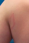
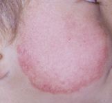
Diagnosis: Erythema migrans/Lyme disease

Annular is derived from the Latin word "annulus," meaning ringed.1 Erythema chronicum migrans appears as ovoid or circular macules/papules or patches/plaques with an erythematous periphery and sometime central clearing. Annular eruptions are common in children, and careful attention to the morphology, course, and when necessary skin biopsy findings will help to distinguish them.
Erythema migrans is the annular erythema that is pathognomonic of Lyme disease. The causative organism Borrelia burgdorferi is introduced by the bite of infected ixodid ticks, and the lesion typically appears seven days later. As the tick feeds, a small inoculum of the spirochete enters the skin, resulting in a subtle small red papule that expands outward, producing a round pink to red patch or plaque. The patch increases in size to 5 to 10 cm over several weeks, with some lesions growing to 50 cm in diameter.2 Some patients will complain of minimal tenderness and/or itching. Multiple lesions can occur due to hematogenous dissemination.
The diagnosis is usually based on clinical findings, history of tick exposure, and history of living in or travel to endemic areas. Serology examination for B burgdorferi antibodies can be used as a diagnostic tool. However, false negative results are common during the first few weeks of infection, and false-positive results are common since routine serology cannot distinguish between active and past infections.3 Doxycycline is the treatment of choice for erythema migrans in patients over eight years of age. Amoxicillin should be used in younger children.
Differential diagnosis
Unlike tinea corporis and erythema multiforme, the edge of erythema migrans remains flat or subtly edematous, blanches with pressure, and is not associated with epidermal changes such as scale, vesicles, crusts, or pustules. While urticaria may share features with erythema migrans, they are transient and rarely last more than 12 hours. Erythema annulare centrifigum tends to be chronic, asymptomatic, and associated with epidermal changes. These annular eruptions may overlap clinically with erythema migrans and will be discussed in detail.
Tinea corporis
Tinea corporis, referred to as "ringworm," is a superficial fungal infection caused by dermatophytes that most commonly infects the hands (tinea manuum), feet (tinea pedis), scalp (tinea capitis), face (tinea faciei), nails (tinea unguium onychomycosis) or groin (tinea cruris).4 The most common organisms associated with tinea corporis in the United States include Trichophyton rubrum, Trichophyton tonsurans, Trichophyton mentagrophytes, and Microsporum canis.

The presence of the characteristic superficial itchy annular scaly plaques should suggest the diagnosis, which can be confirmed by potassium hydroxide preparation and/or fungal culture. Localized lesions respond to topical antifungal medications, but oral antifungal therapy should be considered for widespread lesions, eruptions in difficult-to-reach areas, or persistent infections in the groin, perianal skin, or plantar surfaces of the feet.5
Granuloma annulare
Granuloma annulare (GA) is a common idiopathic inflammatory dermatosis involving the dermis and/or subcutaneous tissue. The localized cutaneous variant of GA is quite often confused with other types of annular eruptions.1,6
Recognize & Refer: Hemangiomas in pediatrics
July 17th 2019Contemporary Pediatrics sits down exclusively with Sheila Fallon Friedlander, MD, a professor dermatology and pediatrics, to discuss the one key condition for which she believes community pediatricians should be especially aware-hemangiomas.