Tularemia in a 4-Year-Old Girl
During spring vacation, a previously healthy 4-year-old girl visited western Nebraska, where she and her family spent time along a river bank in a wooded area. After 4 days, her mother noticed 3 engorged ticks embedded in the child's scalp. The ticks were immediately removed and burned. The child also had a marble-sized swelling on the right side of her neck. Over the next few days, the child had temperatures that spiked to 39.4C (103F), with chills, generalized malaise, and weakness. There was no history of cough, myalgias, or headache.

Figure

Figure
HISTORY
During spring vacation, a previously healthy 4-year-old girl visited western Nebraska, where she and her family spent time along a river bank in a wooded area. After 4 days, her mother noticed 3 engorged ticks embedded in the child's scalp. The ticks were immediately removed and burned. The child also had a marble-sized swelling on the right side of her neck. Over the next few days, the child had temperatures that spiked to 39.4C (103F), with chills, generalized malaise, and weakness. There was no history of cough, myalgias, or headache.
Her pediatrician diagnosed lymphadenitis and prescribed cephalexin. After a few days of therapy, the child was afebrile and felt better. However, the swelling in her neck had increased in size. She also had 3 other swellings on the back of her neck. The patient was referred for further evaluation.
PHYSICAL EXAMINATION
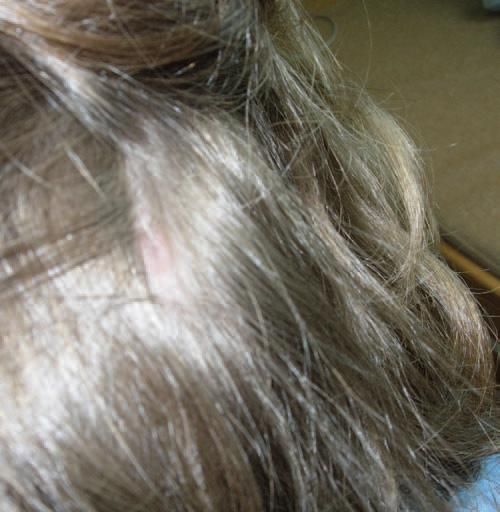
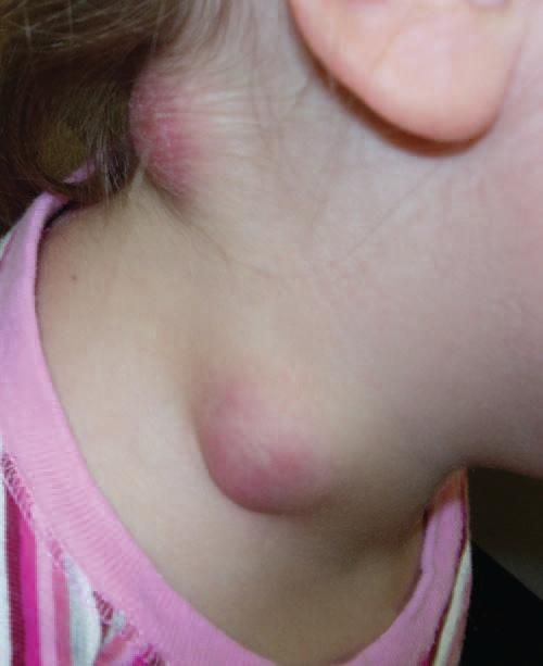
The patient was calm and in no distress. Vital signs were normal. Examination of the head revealed a small scab of 5 5 mm with alopecia over the right occipital region. She had 4 swellings in the posterosuperior cervical area. Two were 3 3-cm, globular, soft, nontender masses with an erythematous hue and slight desquamation on the surface. There were 2 smaller swellings in the occipital region. The remainder of the physical examination findings were unremarkable.
LABORATORY INVESTIGATIONS
Results of a complete blood cell count and comprehensive metabolic profile were normal. Blood cultures remained negative, and a tuberculin skin test result was negative at 48 hours. Gentamicin (2.5 mg/kg every 8 hours) was started.
WHAT'S YOUR DIAGNOSIS?
ANSWER: TULAREMIA
A week after the initial laboratory tests, direct agglutination showed an antibody titer of 1:320 against Francisella tularensis, which confirmed the diagnosis of tularemia. Confirmation of this diagnosis requires a single titer of at least 1:160 by tube agglutination or a 4-fold or greater increase in titers between 2 serum samples obtained at least 2 weeks apart.1
The patient received 1 week of gentamicin therapy, during which 1 node spontaneously drained purulent material. Gram stain of this fluid revealed a Gram-negative rod. Cultures were negative.
The lymphadenitis had decreased significantly at follow-up, and the patient was given oral doxycycline for 2 more weeks. Follow-up serological testing after 3 weeks revealed antibody titers of 1:160. At 4 weeks' follow-up, she had a well-healed scab with no new abscesses.
TULAREMIA
Tick bites are the predominant mode of transmission of tularemia in the continental United States.1 Regional lymphadenopathy after a tick bite is a common presenting feature.2 The diagnosis can be missed if a high index of suspicion is not maintained.
Tularemia is a zoonosis caused by F tularensis, a small, nonmotile, Gram-negative, intracellular coccobacillus. It can be found in several animal hosts, such as rabbits, hares, cats, voles, mice, and many other small mammals and rodents. Humans become infected through bites of infected arthropods-particularly ticks-as well as through contact with infected animals, ingestion of contaminated food or water, and inhalation of aerosolized organisms. With the reduction in hunting and trapping, tick bites have become the most common source of infection in the United States.3 Tularemia is highly infectious; inoculation with as few as 10 organisms can cause disease.2,3
Bacterial exposure through broken skin results in ulceroglandular or glandular disease. Other disease presentations include pleuropneumonitis and oculoglandular, oropharyngeal, and typhoidal tularemia. F tularensis is a potential bioterrorism agent.1,4
The onset of tularemia is usually abrupt with flu-like symptoms, such as fever, headache, chills, rigors, and generalized myalgia. A high index of suspicion is needed because the symptoms are nonspecific and diagnosis can be delayed if a detailed history (including that of animal and insect bites) is not elicited. Regional lymphadenopathy is common in patients infected through the skin. Adenitis is noted usually in the inguinal or femoral nodes because most tick bites occur on the extremities or trunk. In our patient, the swelling in the neck was mistaken for suppurative lymphadenitis secondary to Staphylococcus aureus infection.
When tularemia is suspected, the diagnostic workup should include cultures of blood and sputum (if present) and/or aspirate from a lymph node or abscess. Isolation of F tularensis is difficult because it is a strict aerobic organism and requires cysteine-enriched media for growth.2,3 The diagnosis is confirmed serologically by tube agglutination. A titer of 1:160 is consistent with a recent or past infection and constitutes a presumptive diagnosis. Direct fluorescent antibody or polymerase chain reaction assays, which can be performed on sputum or material from a lesion or abscess, are rapid diagnostic procedures but are not routinely performed.5
Tularemia is treated with gentamicin at 2.5 mg/kg given intravenously or intramuscularly every 8 hours for 10 days.1,3 Other treatment options include streptomycin, doxycycline, chloramphenicol, and ciprofloxacin. Bactericidal drugs are used for 10 days, but bacteriostatic drugs should be used for a minimum of 14 days.1
In our patient's case, the abscess did not respond to gentamicin because it had not been drained. However, after the abscess spontaneously drained, the lesions healed completely with an additional course of doxycycline.
When evaluating patients with neck swelling, tularemia must be included in the differential diagnosis, and a thorough history should be elicited.
References:
- Dennis DT, Inglesby TV, Henderson DA, et al. Tularemia as a biological weapon: medical and public health management. JAMA. 2001;285:2763-2773.
- Spach DH, Liles WC, Campbell GL, et al. Tick-borne diseases in the United States. N Engl J Med. 1993;329:936-947.
- Eliasson H, Broman T, Forsman M, Bäck E. Tularemia: current epidemiology and disease management. Infect Dis Clin North Am. 2006;20:289-311.
- Feldman KA, Enscore RE, Lathrop SL, et al. An outbreak of primary pneumonic tularemia on Martha's Vineyard. N Engl J Med. 2001;345:1601-1606.
- Johansson A, Berglund L, Eriksson U, et al. Comparative analysis of PCR versus culture for diagnosis of ulceroglandular tularemia. J Clin Microbiol. 2000;38:22-26.
Recognize & Refer: Hemangiomas in pediatrics
July 17th 2019Contemporary Pediatrics sits down exclusively with Sheila Fallon Friedlander, MD, a professor dermatology and pediatrics, to discuss the one key condition for which she believes community pediatricians should be especially aware-hemangiomas.