Weeks of weakness, then reddish bumps on prominences
A boy with a history of skin eruption, intermittent fever, and generalized weakness now has red bumps on the bony prominences of his hands, elbows, and knees. What's the diagnosis?
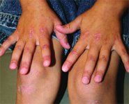
You are asked to see a 6-year-old boy who has a history of a skin eruption, intermittent fever, and generalized weakness. His mother is concerned about increasing puffiness around his eyes and red bumps on the bony prominences of his hands, elbows, and knees (see the photograph).

The history and clinical findings are typical of juvenile dermatomyositis (JDM). Further evaluation-including elevated serum levels of creatine kinase and aldolase, skin biopsy findings consistent with connective tissue disease, and evidence of muscle inflammation on a magnetic resonance imaging (MRI) scan-all confirm your clinical suspicion.
Presentation
JDM is a chronic inflammatory disorder of unknown etiology that primarily affects muscle and skin. The diagnosis requires not only the presence of one of the characteristic skin findings, but also at least three of four of the following features:
The skin manifestations of JDM include the characteristic heliotrope rash of violaceous erythema and edema of the malar and periorbital skin. A scaly, red, psoriasiform eruption may involve the extensor surfaces of knees and elbows. When this eruption appears on the distal interphalangeal joints, the papules and plaques are referred to as Gottron papules.2 Seborrheic-like dermatitis of the scalp is often present in JDM. Nail findings include periungual erythema and nailfold telangiectasias.
Over time, children with JDM may develop chronic skin changes, including atrophy, hyperpigmentation, fibrosis, and telangiectasias. Late in the course, deposition of calcium in skin, muscle, and joints may result in debilitating contractures, painful ulceration, and cellulitis.3
Differential diagnosis
The facial rash of JDM in children can suggest systemic lupus erythematosus; in fact, some of the clinical features of these two connective-tissue diseases overlap. Severe muscle involvement, with evidence of myositis on MRI or muscle biopsy, or both, and elevation of muscle enzymes, is characteristic of JDM and distinguishes the two disorders.
In early JDM, when muscle symptoms are subtle, the psoriasiform eruption on the extensor surfaces of the extremities can be confused with psoriasis.
The diffuse erythema and scaling of the scalp in JDM can mimic seborrheic dermatitis or psoriasis before the other signs and symptoms of JDM appear.
Treatment and course
An oral corticosteroid, 1-2 mg/kg/day, is the initial treatment of choice for most patients with JDM. Although most patients respond to steroid therapy, some children suffer severe complications of the disease, including gastrointestinal tract and skin ulcerations and dystrophic calcification.4 Other immunosuppressive agents, including methotrexate, azathioprine, intravenous immunoglobulin, and cyclosporine, have been used to treat patients who fail to respond to, or experience severe side effects from, a corticosteroid.
Physical therapy is important to promote recovery. Patients should be encouraged to minimize the risk of a photosensitivity reaction by wearing sunscreen and avoiding exposure to intense sunlight. Muscle strength and enzyme levels should be monitored continuously for signs of relapse.
The prognosis for children who have JDM has improved with the advent of the use of corticosteroids. Early, aggressive treatment has been demonstrated to improve outcome and decrease the incidence of calcinosis.5
REFERENCES
1. Bohan A, Peter JB: Polymyositis and dermatomyositis (first of two parts). N Eng J Med 1975;292:344
2. Sontheimer RD: Dermatomyositis: An overview of recent progress with emphasis on dermatologic aspects. Dermatol Clin 2002;20:387
3. Santmyire-Rosenberger B, Dugan EM: Skin involvement in dermatomyositis. Curr Opin Rheumatol 2003;15:714
4. Huber AM, Lang B, LeBlanc CM, et al: Medium- and long-term functional outcomes in a multicenter cohort of children with juvenile dermatomyositis. Arthritis Rheum 2000;43:541
5. Fisler RE, LIang MG, Fuhlbrigge RC, et al: Aggressive treatment of juvenile dermatomyositis results in improved outcome and decreased incidence of calcinosis. J Am Acad Dermatol 2002;47:505
Please see Dr. Cohen's Web site, http://www.dermatlas.org/, for additional images
Recognize & Refer: Hemangiomas in pediatrics
July 17th 2019Contemporary Pediatrics sits down exclusively with Sheila Fallon Friedlander, MD, a professor dermatology and pediatrics, to discuss the one key condition for which she believes community pediatricians should be especially aware-hemangiomas.