Acute Hemorrhagic Edema of Infancy in an 11-Month-Old Boy
Acute hemorrhagic edema of infancy is a relatively uncommon form of leukocytoclastic vasculitis. Henoch-Schönlein purpura is the primary differential diagnosis.
An 11-month-old boy presented with mild fever and rhinorrhea of several day’s duration. The previous day the child’s mother noted what appeared to be insect bites on the patient's left leg. She became concerned when she noticed his left foot had become swollen. She took the child to a local emergency department where he was given a diagnosis of early-stage Henoch-Schnlein purpura. The mother was instructed to take the child to his regular pediatrician for further examination.

Figure 1
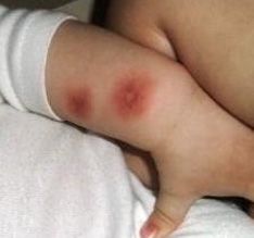
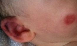
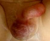
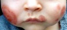
Physical examination revealed a well-appearing, interactive child. He was alert, distractible, and in no acute distress. Several erythematous round, flat-topped papules, less than 0.5 cm in diameter, were noted on his left leg and torso. Moderate, non-pitting edema was noted over his left foot. A pale dusky discoloration developed in the foot over the next several hours and the edema spread proximally to involve most of the left lower leg. The next day, additional lesions were noted on the left leg and trunk and the eruptions had spread to the patient’s arms, ears, face, and genitalia (Figures 1-4). Based on the dramatic onset of signs and symptoms and the child’s relatively benign clinical presentation, a diagnosis of acute hemorrhagic edema of infancy (AHEI) was made. No treatment was prescribed but the parents photographed the skin lesions daily and sent them via Internet to the treating physician’s office for review. The eruptions and edema were completely cleared within 10 days.


Discussion
Acute hemorrhagic edema of infancy (AHEI) is a relatively uncommon form of leukocytoclastic vasculitis that typically presents in children from age 4 months to 2 years. Approximately 100 cases have been reported since the disease was first described in the early 1900s. The small number of cases suggests true low disease incidence but may also reflect underdiagnosis or confusion with other disease entities. Onset is often dramatic with petechiae, ecchymoses, and annular, nummular or targetoid purpuric lesions and usually involve the extremities, face, or ears. The edema is often asymmetric and begins distally and can progress proximally. Extracutaneous involvement is rare, occurring in fewer than 10% of cases, and includes glomerulonephritis, abdominal pain, and arthralgia.
Until recently, most reports of AHEI occurred in the European literature under the terms Finkelstein’s disease, Seidlmayer syndrome, infantile post-infectious iris-like purpura and oedema, and purpura en cocarde avec oedema.
Although the precise cause is unknown, there are documented associations of AHEI with prior viral infection (upper respiratory tract infection, otitis media, viral conjunctivitis), bacterial infection (streptococcal or staphylococcal pharyngitis, pulmonary tuberculosis, pneumonia, urinary tract infection), vaccination, or use of medication (penicillin, cephalosporin, trimethoprim-sulfamethoxazole, or acetaminophen).
AHEI was once considered a variant of Henoch-Schnlein purpura (HSP) but today is recognized as a distinct entity. HSP, however, is still the primary differential diagnosis to be considered. Characteristics that distinguish the conditions include age of onset, which is younger for AHEI (approximately 4 to 24 months) than for HSP (peak age of 4 to 7 years). The systemic complications often seen in HSP (ie, arthralgia, gastrointestinal hemorrhage, nephritis) are extremely rare in AHEI. Clinically, HSP manifests with raised purpura on the extensor surfaces of the legs and on the buttocks. The larger erythematous-edematous purpura and the ecchymoses of AHEI are found on the face and extremities, with or without mucosal involvement; in our case the genitals were also affected. Other diagnoses to be excluded are meningococcemia, erythema multiforme, urticarial vasculitis, Kawasaki disease, and child abuse.
It is often noted that the most remarkable feature of AHEI is the contrast between the intensity of the cutaneous signs, which are typical and unmistakable, and the patient’s otherwise overall good health.
AHEI usually resolves spontaneously within 1 to 3 weeks. Antibiotics are recommended when there is evidence of infection, otherwise treatment is supportive. Systemic corticosteroids and antihistamines have been used in the past but have not been shown to alter the course of disease. There is some evidence that antihistamines may prompt more rapid healing. Ibuprofen and acetaminophen may be used to relieve fever, systemic symptoms, and discomfort but are not specifically indicated.
In this case, the disease ran a typical course and the child required no supportive care. This case also demonstrates the use of technology to monitor a cutaneous disease process. The authors believe this is the first case reported in the literature of a dermatologic disease monitored and treated completely through the exchange of photographs taken in the patient’s home and transmitted for assessment via the Internet. It is essential to differentiate this remote visual monitoring process from the use of patented mobile applications designed to analyze and assign risk to dermatologic lesions. The diagnosis of our patient was made in the clinic and initial therapeutic decisions were made at that time. Personal technology that enhances patient education and can support disease self-management is certainly valuable but self-diagnosis of cutaneous lesions or of any medical condition is highly discouraged.
Suggested Readings on AHEI
• Emerich PS, Prebianchi PA, Motta LL, Lucas EA, Ferreira LM. Acute hemorrhagic edema of infancy: report of three cases. An Bras Dermatol. 2011;86:1181-1184.
• Fotis L, Nikorelou S, Lariou MS, Delis D, Stamoyannou L. Acute hemorrhagic edema of infancy: a frightening but benign disease. Clin Pediatr (Phila). 2012;51:391-393.
• Karremann M, Jordan AJ, Bell N, et al. Acute hemorrhagic edema of infancy: report of 4 cases and review of the current literature. Clin Pediatr (Phila). 2009;48:323-326.
• Acun C, Ustundag G, Sogut A, et al. Visual diagnosis: a child who has acute onset of unusual skin lesions and edema. Pediatr Rev. 2006;27:e71-e74.
• Cicero M. Rash decisions: acute hemorrhagic edema of infancy in a 7-month-old boy. Pediatr Emerg Care. 2008;24:501-502.
• AlSufyani MA. Acute hemorrhagic edema of infancy: unusual scarring and review of the English language literature. Int J Dermatol. 2009;48:617-622.
• Legrain V, Lejean S, Taieb A, et al. Infantile acute hemorrhagic edema of the skin: study of ten cases. J Am Acad Dermatol. 1991;24:17-22.
• Hamilton AD, Brady RR. Medical professional involvement in smartphone 'apps' in dermatology. Br J Dermatol. 2012;167:220-221.
• Mosa AS, Yoo I, Sheets L.A Systematic Review of Healthcare Applications for Smartphones. BMC Med Inform Decis Mak. 2012;12:67