The care of lines and tubes in children
The implantation of fabricated devices has become part of the standard therapeutic approach for many neonatal and pediatric disorders.

Key Points
The implantation of fabricated devices has become part of the standard therapeutic approach for many neonatal and pediatric disorders. Catheters, ostomy tubes, and other apparatus provide access to internal organ systems, thereby facilitating treatment objectives such as drug delivery, nutritional support, and control of secretions. This equipment can be associated with significant morbidity that can adversely affect outcomes. Clinicians taking care of patients with these devices should have a basic understanding of the indications, implantation procedures, risks, and potential complications with their use.
Central venous catheters
Central venous catheters (CVCs) are commonly used in children with nonfunctioning gastrointestinal tracts who require long-term intravenous nutritional therapy or in children who require long-term venous access for pharmacologic treatment such as chemotherapy or chronic transfusion therapy. Complications can occur in 5% to 20% of patients and include fracture with leakage, occlusion with embolization, migration, dislodgement, and infection.1-3
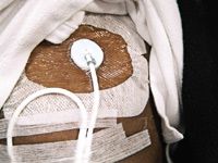
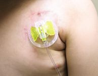
Peripherally inserted central catheter (PICC). These are long, thin catheters that are inserted into a peripheral vein with the distal tip positioned in a central vein. These catheters have several advantages over the tunneled and implanted devices. They may be inserted at the bedside with relative ease; they can be used for outpatient therapy; and they have been shown to be safe in children. Because PICC lines do not require surgery for removal, they are easily removed in an outpatient setting when the child no longer requires them. These tubes are indicated when effective short- and intermediate-term intravenous (IV) access is necessary, such as with IV antibiotic administration for serious infections. These lines confer a low risk of major hemorrhage and no risk of pneumothorax.4-8
A major problem is catheter malfunction from occlusion, infection, venous thrombosis, and fracture, which can occur in 10% of catheters.3,4 Children with catheter malfunction usually present with pain and tenderness at the site. Associated extremity edema or signs of infection, such as erythema or purulence, are indications for prompt removal. Occlusion or breakage of these small catheters cannot be salvaged and are also indications for elective removal. Complications associated with insertion and failures of these catheters have been reduced by the development of institutional PICC teams that are skilled in their management. In general, PICC lines can remain in place for 4 to 6 weeks. Their average lifespan is approximately 1 month, and they have an infection rate of 0.8 episodes per 1,000 catheter days.4,6,8
Externalized tunneled CVCs. These include the Groshong, Hickman, or Broviac catheters. They are available in various sizes and can be single or double lumen.2-4 Like PICC lines, they are used when long-term IV access is necessary. However, the average lifespan for these catheters is 6 to 12 months, and in many cases, a well-cared-for tunneled CVC can stay in place for years. These catheters have a lower incidence of infection and complication than PICC lines, making them more suitable in children who are immunosuppressed from a chronic medical or cancer condition. The most common indication is for IV therapy related to malignancy. They are made of a silicone elastomer, which causes less trauma to the surrounding tissues of the child on insertion and offers some resistance to infection and thrombus formation compared with nontunneled CVCs. The 2.9 mm- to 9 mm-diameter catheters are most commonly used in the pediatric population. These catheters have a polyester fabric cuff that is positioned subcutaneously at or just proximal to the skin exit site. The cuff stimulates a fibrous reaction that serves to anchor the catheter, reducing the likelihood of inadvertent displacement or migration, and serves as a mechanical barrier to exit-site and tunnel infections.3
Typically, these catheters are placed percutaneously, except in the very small infant, in which case the external or internal jugular vein is accessed via a surgical cut down. The most common sites for percutaneous access are the subclavian vein and external and internal jugular veins. The catheter is then tunneled from the venous access site through the subcutaneous tissues to a remote site, usually on the anterior chest.

Catheter malfunction from dislodgement, breakage, occlusion, infection, and venous thrombosis is common.3 Parents of children presenting to a pediatrician's office with a nonfunctioning or dislodged catheter report an inability to flush the catheter or pain with infusion. However, most of these catheters can be salvaged. It is recommended that these children be referred to the hospital because many of the strategies for salvage require inpatient care. It is important to determine and document in the child's record the specific size catheter used because this information is vital to repair the catheter if the external portion fractures. The repair kits are specific to the size of catheter (ie, a 4-mm catheter will require a 4-mm repair kit). In a cooperative child with a clear fracture of the externalized portion, many pediatricians could repair the catheter in the office.
