Dislodged Suture Anchor After Gastrostomy

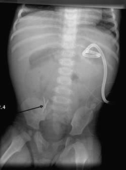
A 17-month-old boy with a history of gastroschisis and functional short-gut syndrome was admitted for treatment of central line sepsis with intravenous antibiotics. During therapy, non-bilious, non-bloody vomit and abdominal distention developed. X-ray films of the abdomen revealed 2 radio-opaque needle-like objects at the ileocecal area (Figure 1).
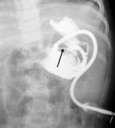
The radiologist was able to compare this image with an abdominal film taken 2 months earlier during image-guided percutaneous insertion of the gastrostomy tube (Figure 2). The arrow in that image points to 1 of 2 retention tacks (gastrointestinal suture anchor) placed during that procedure. The second retention tack is overshadowed by the contrast.
The GI suture anchor secures the anterior wall of the stomach to the abdominal wall before interventional catheters are inserted. Details of the procedure are well illustrated in the literature.1 Complications from

gastrostomy and the tubes are well known2 but we are not aware of any documented problems caused by the suture anchor. This was an incidental finding, unrelated to the patient’s abdominal problems.
Conceivably, suture anchors could become lodged and retained in the gut of a child with strictures or poor motility. In time, a suture anchor usually dislodges from the inside of the stomach and is passed in the stool.
This patient was discharged home; follow-up abdominal x-rays showed no retention tack in the intestine, which was (presumably) evacuated in the stool.