- Consultant for Pediatricians Vol 6 No 8
- Volume 6
- Issue 8
Linear IgA Dermatosis (Childhood Type) and Fixed Drug Eruption
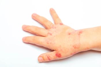
This 6-year-old child was brought to the emergency department by her parents after these blisters suddenly appeared on her skin. She otherwise appeared to be well and was not febrile.
Case 1:
This 6-year-old child was brought to the emergency department by her parents after these blisters suddenly appeared on her skin. She otherwise appeared to be well and was not febrile.
Your clinical examination can easily classify her condition and allow you to be very certain as to its severity. Should you worry about this child? What's the cause?
Case 2:
This boy presented with an inflammatory plaque on his skin that was intensely "itchy and burny." It was the third time that the lesion had occurred in the same location, and it was always associated with sore knees following gym class.
What is the cause?
Case 1: Linear IgA dermatosis-childhood type
The development of blisters on a child's skin is one of the most worrisome events in pediatric dermatology. The diagnosis may be as simple as an insect bite reaction--or as life-threatening as toxic epidermal necrolysis.
The morphology of blisters in a child can help you quickly ascertain the seriousness of the condition. Blistering diseases are categorized by the position of the blister within the skin itself. For example, blisters associated with bullous impetigo are very flaccid or may simply be represented by an erosion because the blister cavity is through the stratum corneum, which is the most superficial aspect of the skin. In contrast, the blister cavity in bullous pemphigoid is through the basement membrane zone at the dermoepidermal junction: it produces a tense blister that is not easily brushed from the skin.
I also look for blisters on noninflamed skin. Their presence usually indicates that the condition is systemic.
This young girl presented with widespread tense blisters on inflamed and noninflamed skin (A). These lesions had a predilection for the face, lower trunk, and genitalia. A ring of blisters on the trunk (called the "string of pearls" sign) was also present. There were no lesions on this child's mucous membranes.
Her main symptoms were mild itch, discomfort, and inflammation around her genitalia. Her clinical presentation is most consistent with the diagnosis of chronic bullous dermatosis of childhood, now referred to as linear IgA dermatosis-childhood type.
The evaluation of a bullous disease of the skin invariably requires a biopsy specimen for both routine and immunofluorescence staining. Ideally, the specimen is taken from the inflammatory border of the blister and the edge of the blister cavity. Cells at these sites can define the histological nature of the process. Direct immunofluorescence further classifies the nature of the condition.
In linear IgA dermatosis-childhood type, the condition is defined by the immunofluorescence. A deposition of IgA in a linear pattern along the basement membrane zone (B) is the defining characteristic of this condition and is present in all affected persons. Routine histological evaluation (C) shows a subepidermal blister with dermal edema. Neutrophils and eosinophils invade the dermis at the blister edge.
This condition is one of early childhood: it usually occurs within the first decade. It may resolve spontaneously within a few years; virtually no patients suffer past puberty. Systemic therapy with dapsone (Avlosulfone) is the treatment of choice, and the condition is almost always quickly responsive.
Case 2: Fixed drug eruption
A fixed drug eruption (FDE) has developed in response to the NSAID this boy had been taking to alleviate the discomfort in his knees. The most telling clinical clue is the recurrence of the lesions in the same location with each appearance.
FDE is a peculiar reaction to the ingestion of a variety of chemicals--including drugs. Although it is unusual, it is easily recognizable by its characteristics. Once you have seen one, it is easy to remember.
An FDE always occurs at the same location, although other sites may be recruited with each subsequent reaction. The reaction is usually preceded by an itch or burning at the site; this is followed by the development of an intense erythematous and edematous plaque that becomes dusky red or violaceous and which may blister. The genitalia, lips, hands, and feet are the sites of predilection. If there are multiple lesions, they will all occur at the same time; their appearance and symptoms will also be similar. The first occurrence begins within 1 to 2 weeks of the drug ingestion, but subsequent reactions usually occur within 24 hours of medication exposure.
The lesions begin to fade over 1 to 2 weeks. The defining feature of the reaction is the dark blue-black hyperpigmentation, which occurs at all sites of reaction and which marks the sites where the lesions will recur.
The drugs most commonly associated with FDEs are trimethoprim/sulfamethoxazole, NSAIDs (ibuprofen), paracetamol (acetaminophen), antipyretics, and tetracyclines. There are lists of medications and foods known to be associated with FDE. You can confirm that a drug is implicated when a second identical reaction occurs after a repeated ingestion.
Once you have made the diagnosis--which may occasionally require a biopsy--a detailed medication history that includes prescription and over-the-counter drugs needs to be completed. Agents that are taken intermittently are the most likely culprits.
Unfortunately, avoidance is the only successful course of action. Once the reaction has begun, it has been my experience that nothing reverses or attenuates it. *
Articles in this issue
over 18 years ago
Asymmetric Crying Faciesover 18 years ago
Cystic Hygroma in a 1-Year-Old Girlover 18 years ago
Hypertransaminasemia: A Diagnostic Dilemmaover 18 years ago
Toxic Epidermal Necrolysis Secondary to Anticonvulsant Medicationover 18 years ago
TTN--A Benign Condition or Precursor to a Chronic Illness?over 18 years ago
Winter 1979over 18 years ago
Itchy Spreading Rash on Teen's Cheekover 18 years ago
Dental Disease: REFERENCESover 18 years ago
Feedback on Snake Bite: A Small Puncture Can Create a Large ProblemNewsletter
Access practical, evidence-based guidance to support better care for our youngest patients. Join our email list for the latest clinical updates.


