


Novel genetic therapies for sickle cell disease and β-thalassemia
As safe and effective gene-editing and gene-replacement therapies become more attainable, developing the delivery systems that ensure these treatments are equitably available will be equally essential.


Novel genetic therapies for sickle cell disease and β-thalassemia | Image Credit: © 736405972 - © 736405972 - stock.adobe.com.
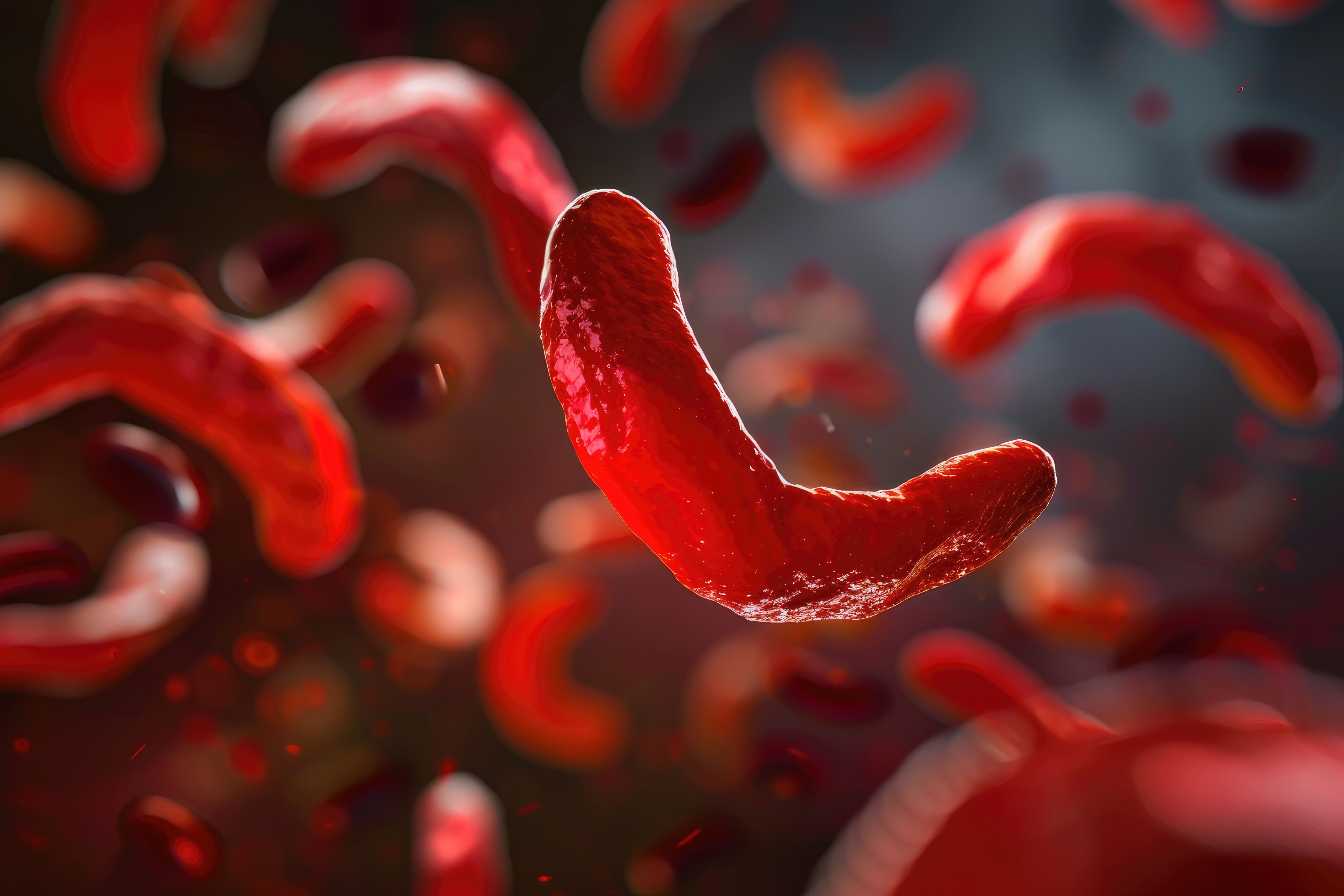
Sickle cell disease (SCD) and β-thalassemia are hemoglobinopathies caused by pathogenic variants in HBB, the gene-encoding β-hemoglobin. SCD is caused by the presence of the p.Glu6Val pathogenic variant in HBB (termed HbS), compounded with a second HBB pathogenic variant.1 Deoxygenation leads to HbS polymerization, causing sickling of erythrocytes, microvascular occlusion, and the well-known sequelae of SCD. β-thalassemia major is caused by biallelic pathogenic variants in HBB that result in absence of β-hemoglobin synthesis (Hb β0/β0), causing severe chronic anemia and transfusion dependence (transfusion-dependent β-thalassemia [TDT]).2
On December 8, 2023, the FDA approved 2 cell-based gene therapies, exagamglogene autotemcel (Casgevy; Vertex and CRISPR Therapeutics) and lovotibeglogene autotemcel (Lyfgenia; Bluebird Bio, Inc), for the treatment of sickle cell disease (SCD) in patients 12 years and older. Exagamglogene autotemcel has also been investigated as gene therapy for TDT. While these therapies work by distinct molecular mechanisms, both had promising safety and efficacy data from multicenter trials in adolescents and adults with SCD. These are the first gene therapies to be FDA approved for SCD, and exagamglogene autotemcel is the first gene-editing therapy to treat human disease. Both therapies utilize ex vivo gene therapy of patient-derived CD34+ hematopoietic stem cells (HPSCs), which are then transplanted to the patient.
Exagamglogene autotemcel (Casgevy)
Exagamglogene autotemcel, developed by Vertex and CRISPR Therapeutics, uses clustered regularly interspaced short palindromic repeats (CRISPR)–Cas9 gene editing to disrupt the erythroid-specific enhancer region of BCL11A, thereby increasing fetal hemoglobin (HbF) synthesis. HbF is protective in SCD by decreasing rates of erythrocyte polymerization and associated morbidity and mortality.3,4 The biological switch from HbF to adult hemoglobin (predominantly HbA) occurs during the neonatal period, and HbF is rapidly downregulated after birth as adult Hb becomes expressed.3,5 Approximately 50% of HbF silencing in adults occurs via the transcription factor BCL11A, the genetic target which exagamglogene autotemcel disrupts.
The treatment process starts with apheresis of CD34+ HPSCs from the patient’s bone marrow. These cells are then electroporated to allow for incorporation of the CRISPR-Cas9 gene editing machinery. This allows for a specific guide RNA to direct Cas9 to the BCL11A enhancer region, where the Cas9 endonuclease catalyzes a double-stranded break. Nonhomologous end-joining mediated by the cell’s intrinsic DNA repair machinery then reanneals the break, a process accompanied by the introduction of small deletions or insertions (indels) at the repair site. These indels ablate the erythrocyte enhancer region of BCL11A, leading to reduced expression of BCL11A in erythroid lineage cells and increased endogenous production of HbF. Hematopoetic stem cell transplantation (HSCT) of these cells requires busulfan myeloablation, followed by a single intravenous infusion of the gene-edited HPSCs.
Preclinical studies of exagamglogene autotemcel using CD34+ HPSCs from healthy donors showed approximately 80% editing efficiency in all subpopulations of CD34+ cells, an increase in HbF to 29.0% in edited erythroid cells (10.5% in unedited cells), and undetectable off-target editing at over 200 candidate sites.5
Exagamglogene autotemcel was investigated in a phase 3, single-dose, open-label study (NCT05477563) in participants with TDT and severe SCD. Results from the first 2 treated individuals with SCD and TDT showed persistence of a high frequency of edited alleles 12 months after transplantation in both peripheral blood and bone marrow, along with improved anemia and increased circulating erythrocytes expressing HbF over the study duration. Both patients had marked improvements in their symptoms following treatment, including no further dependence on red blood cell transfusions in either patient, and no vaso-occlusive crises or hospitalizations in the patient with SCD.5,6
At the time of regulatory agency approval, 28 of 29 enrolled patients with SCD endorsed complete relief of debilitating symptoms. Thirty-nine of the 42 patients with TDT no longer required red blood cell transfusions for more than 1 year, and the remaining 3 patients’ transfusion needs were reduced by 70%. While a 15-year clinical follow-up is still underway, these promising results provided sufficient evidence of safety and efficacy to US and European regulatory agencies for clinical approval.
Lovotibeglogene autotemcel (Lyfgenia)
Lovotibeglogene autotemcel was developed by Bluebird Bio and uses a viral vector gene therapy approach to express the HbAT87Q variant in SCD patient erythrocytes. This variant of adult hemoglobin, with a threonine to glutamine substitution at residue 87, sterically inhibits polymerization of HbS and produces an anti-sickling hemoglobin. Upon apheresis of CD34+ HPSCs from patient bone marrow, cells are transduced with a lentivirus vector encoding the complementary DNA of the modified HBB with the HbAT87Q substitution. HSCT of the genetically modified HPSCs is performed following myeloablation with busulfan.
This treatment was investigated in a phase 1/2, open-label, single-dose clinical trial in participants with SCD who failed hydroxyurea treatment and had active disease (NCT02140554).7,8 At 24 months following infusion of lentiviral-transduced HPSCs, approximately 85% of erythrocytes were estimated to contain HbAT87Q. Treatment was associated with reduced hemolysis and improvement in anemia, with median hemoglobin levels above 11 g/dL (from 8.5 g/dL) sustained for up to 36 months after infusion. Among 25 patients evaluated, 3 (12%) had vaso-occlusive events after infusion of therapy, but all had resolution of severe vaso-occlusive crises (defined as an event requiring hospitalization, presentation to an emergency department, or priapism).7-9
Risks
Exagamglogene autotemcel and lovotibeglogene autotemcel show auspicious potential as disease-modifying therapies for sickle cell disease and β-thalassemias, and their development and regulatory approval is a major achievement for the gene therapy and gene-editing fields. For both strategies, several important clinical questions remain including the clinical risks of myeloablation (infections, infertility), long-term clinical durability, unclear off-target effects from gene editing in exagamglogene autotemcel, and unclear risks of developing myelodysplastic syndromes from lentiviral insertion in lovotibeglogene autotemcel. Of note, among participants treated with lovotibeglogene autotemcel, 2 patients developed acute myeloid leukemia after infusion, although there was no direct evidence of insertional oncogenesis.7,10
Conclusion
Access to therapy is also a considerable limitation, with exagamglogene autotemcel estimated to cost over $2 million per patient and HSCT requiring specialized centers. Given the high costs, this therapy is unattainable for the many patients with SCD or TDT who reside in resource-poor countries. As safe and effective gene editing and gene-replacement therapies become more attainable, developing the delivery systems that ensure these treatments are equitably available will be equally essential.
References:
1. Kavanagh PL, Fasipe TA, Wun T. Sickle cell disease: a review. JAMA. 2022;328(1):57-68. doi:10.1001/jama.2022.10233
2. Langer, AL. In: Adam MP, ed. GeneReviews. University of Washington, Seattle, WA; 1993
3. Paschoudi K, Yannaki E, Psatha N. Precision editing as a therapeutic approach for β-hemoglobinopathies. Int J Mol Sci. 2023;24(11):9527. doi:10.3390/ijms24119527
4. Platt OS, Brambilla DJ, Rosse WF, et al. Mortality in sickle cell disease: life expectancy and risk factors for early death. N Engl J Med. 1994;330(23):1639-1644. doi:10.1056/NEJM199406093302303
5. Frangoul H, Altshuler D, Cappellini MD, et al. CRISPR-Cas9 gene editing for sickle cell disease and β-thalassemia. N Engl J Med. 2021;384(3):252-260. doi:10.1056/NEJMoa2031054
6. Pollock G, Negre O, Ribeil JA. Gene-addition/editing therapy in sickle cell disease. Presse Med. 2023;52(4):104214. doi:10.1016/j.lpm.2023.104214
7. Kanter J, Walters MC, Krishnamurti L, et al. Biologic and clinical efficacy of lentiglobin for sickle cell disease. N Engl J Med. 2022;386(7):617-628. doi:10.1056/NEJMoa2117175
8. Kanter J, Thompson AA, Pierciey FJ Jr, et al. Lovo-cel gene therapy for sickle cell disease: treatment process evolution and outcomes in the initial groups of the HGB-206 study. Am J Hematol. 2023;98(1):11-22. doi:10.1002/ajh.26741
9. Crossley M, Christakopoulos GE, Weiss MJ. Effective therapies for sickle cell disease: are we there yet? Trends Genet. 2022;38(12):1284-1298. doi:10.1016/j.tig.2022.07.003
10. Goyal S, Tisdale J, Schmidt M, et al. Acute myeloid leukemia case after gene therapy for sickle cell disease. N Engl J Med. 2022;386(2):138-147. doi:10.1056/NEJMoa2109167


")

Omalizumab outperforms oral immunotherapy in treating multi-food allergy
March 27th 2025A new clinical trial has found that omalizumab (Xolair; Genetech, Novartis) is more effective than oral immunotherapy (OIT) in treating multi-food allergy in individuals with severe allergic reactions to small amounts of common food allergens.