Orbital Abscess
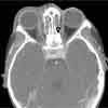
A 4-year-old African American boy presented with his second episode of orbital cellulitis in 9 months. William Demshok, PA-C, of Bascom Palmer Eye Institute, University of Miami, reports that a CT scan showed an abscess in the ethmoidal sinus with extension into the left orbit that impinges on the medial rectus muscle.

A 4-year-old African American boy presented with his second episode of orbital cellulitis in 9 months. William Demshok, PA-C, of Bascom Palmer Eye Institute, University of Miami, reports that a CT scan showed an abscess in the ethmoidal sinus with extension into the left orbit that impinges on the medial rectus muscle.
Most cases of orbital cellulitis are caused by sinusitis, followed by eyelid or facial infection, foreign body, and hematogenous disorder.1,2Staphylococcus and Streptococcus species are the usual causative organisms in adults and children; Haemophilus influenzae has become a less common cause in children with the use of the H influenzae type b vaccine.3,4Pseudomonas species, Escherichia coli, and fungi are less common causes.
All children with suspected orbital cellulitis require a CT scan to investigate the extent of involvement. Intravenous antibiotic therapy, with either ampicillin/sulbactam or ceftriaxone and vancomycin, is also required. Surgical sinus drainage is indicated in about half of cases in children and may be warranted in as many as 90% of cases in adults.1 The differential diagnosis includes trauma with or without retrobulbar hemorrhage, tumor, allergic reaction, insect bite, and herpes zoster.1,5 As always, a thorough history taking is extremely helpful.
Because this child's abscess extended into the left orbit, surgical debridement of the ethmoid sinus was performed. Clindamycin and cefuroxime were given initially and later replaced with ampicillin/sulbactam, as directed by the otorhinolarynology service. Cultures of the drained sinus fluid grew Lactobacillus species and mixed respiratory flora. The patient recovered well, and the cellulitis has not recurred.
References:
REFERENCES:
1.
Kunimoto DY, Kanitkar KD, Makar M, eds.
The Wills Eye Manual: Office and Emergency Room Diagnosis and Treatment of Eye Disease.
4th ed. Philadelphia: Lippincott Williams & Wilkins; 2005:131-133.
2.
Nageswaran S, Woods CR, Benjamin DK Jr, et al. Orbital cellulitis in children.
Pediatr Infect Dis J.
2006;25:695-699.
3.
Ambati BK, Ambati J, Azar N, et al. Periorbital and orbital cellulitis before and after the advent of
Haemophilus influenzae
type B vaccination.
Ophthalmology.
2000;107:1450-1453.
4.
Donahue SP, Schwartz G. Preseptal and orbital cellulitis in childhood. A changing microbiologic spectrum.
Ophthalmology.
1998;105:1902-1905.
5.
Yanoff M, Duker JS.
Ophthalmology.
2nd ed. St Louis: Elsevier; 2004.