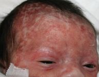
Papules and plaques, on face and scalp, in an otherwise normal newborn
A newborn girl with skin eruption involving the face and scalp, and no adenopathy or organomegaly
DR. COHEN, who serves as section editor for Dermatology: What's your Dx?, is director, Pediatric Dermatology and Cutaneous Laser Center, and professor of dermatology and pediatrics at Johns Hopkins University School of Medicine. He is a contributing editor for Contemporary Pediatrics. He has nothing to disclose in regard to affiliations with, or financial interests in, any organization that may have an interest in any part of this article.

Diagnosis: Neonatal lupus erythematosus
Epidemiology
Neonatal lupus erythematosus is an autoimmune disorder triggered by transplacental maternal antibodies.1-6 Although NLE reportedly occurs in approximately 1 in 12,500 infants, the incidence is probably higher because babies with asymptomatic (undetected) cutaneous disease may go undiagnosed. Among reported cases, more than one half had congenital heart block; 30% to 40% had skin disease; and 10% had both findings. Hematologic involvement and hepatic involvement are less common and usually asymptomatic.
Skin findings
Cutaneous lesions may be present at birth but more commonly develop at several weeks or several months of age-often, following exposure to the ultraviolet (UV) component of sunlight.1-6 The head and neck (especially the scalp) and periorbital and malar areas are most often affected. Discrete and confluent red-to-violaceous, edematous, urticarial papules and plaques usually develop fine overlying scale and dusky central crusting. Lesions heal over six to nine months, often leaving telangiectasias, hypopigmentation or hyperpigmentation, and subtle atrophy. In some patients without systemic findings and subtle early lesions, the earliest manifestations of NLE may be telangiectasias or other late findings, or both.
Extracutaneous findings
Irreversible congenital heart block is the most commonly reported extracutaneous finding; it is usually detected as fetal bradycardia before 30 weeks' gestation. Insertion of a pacemaker in the neonatal period may be required. Other conduction abnormalities and cardiac anomalies have been reported occasionally.
Thrombocytopenia, usually transient, occurs in 10% to 20% of patients. Transient leukopenia, aplastic anemia, and hemolytic anemia may also develop.
Hepatosplenomegaly resulting from extramedullary hematopoiesis may be common, but elevation of liver enzymes is seen in only 10% of cases.
Pathogenesis
The pathogenesis of NLE is not fully understood, but mounting evidence demonstrates a causative role for anti-Ro antibodies.1-6 These antibodies are a marker of disease activity; most symptoms resolve as antibodies disappear by 5 or 6 months of age. Regrettably, damage to the cardiac conduction system and cutaneous scarring are irreversible: Investigators have demonstrated that anti-Ro as well as anti-La and U1RNP antibodies bind to basal keratinocytes and the cardiac conduction system. Binding of these antibodies in skin is potentiated by exposure to UV light.
Making the diagnosis
Neonatal lupus erythematosus should be considered in any child who has congenital heart block or a urticarial annular skin eruption. Development of cutaneous atrophy, telangiectasias, or unusual angiomas during the first year of life may also suggest this diagnosis; a positive test for anti-Ro is diagnostic in this setting. Biopsy of early skin lesions demonstrates characteristic vacuolar changes in basal keratinocytes and a perivascular lymphocytic dermal infiltrate. Direct immunofluorescence testing shows a pattern of speckled keratinocytes typical of NLE and subacute cutaneous lupus erythematosus.
The differential diagnosis of the acute eruption includes urticaria, erythema multiforme, morbilliform drug reaction, viral exanthema, early syphilis, and erythema annulare centrifugum. The course of skin lesions, findings of biopsy, and presence of anti-Ro confirm the clinical diagnosis.
What management is needed?
Cardiac complications should be managed with referral to a pediatric cardiologist. Most other findings are transient, however, and require monitoring but no intervention. Parents should be advised to provide careful sun protection to minimize exacerbation of skin lesions and eventual scarring. Telangiectasias, dyspigmentation, and atrophy may improve with cutaneous laser therapy.
Having "the talk" with teen patients
June 17th 2022A visit with a pediatric clinician is an ideal time to ensure that a teenager knows the correct information, has the opportunity to make certain contraceptive choices, and instill the knowledge that the pediatric office is a safe place to come for help.
Artificial intelligence improves congenital heart defect detection on prenatal ultrasounds
January 31st 2025AI-assisted software improves clinicians' detection of congenital heart defects in prenatal ultrasounds, enhancing accuracy, confidence, and speed, according to a study presented at SMFM's Annual Pregnancy Meeting.