- Consultant for Pediatricians Vol 5 No 9
- Volume 5
- Issue 9
Photoclinic: Bilateral Epiblepharon
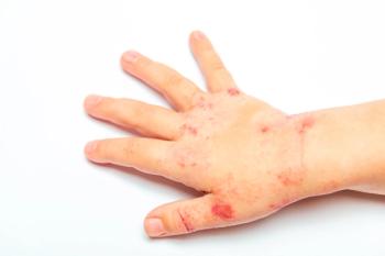
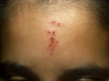
Two children--one with a history of infection, the other with a history of an allergic reaction--were noted to have postinflammatory hyperpigmentation.
Two children--one with a history of infection, the other with a history of an allergic reaction--were noted to have postinflammatory hyperpigmentation.
Photo A shows a 6-year-old girl with 2 hyperpigmented areas, 1 on the right anterior chest and 1 on the upper arm. The previous month, she had impetigo and was treated with oral cephalexin and topical mupirocin ointment. Photo B shows a 14-year-old boy with areas of light tan hyperpigmentation on the neck. At 3 years of age, he had poison ivy dermatitis that resulted in thick black scabs, which eventually peeled away from the edges over several weeks. The resultant hyperpigmentation has remained unchanged for the past 11 years.
Robert P. Blereau, MD, of Morgan City, La, writes that inflammation from allergic or infectious causes can result in hypopigmentation or hyperpigmentation of the involved area. Melanin hyperpigmentation or melanoderma may be classified as follows1:
Chloasma
Incontinentia pigmenti
Secondary to skin diseases (eg, tinea versicolor, fixed drug eruption)
Secondary to external agents (eg, UV light, photosensitizing chemicals)
Secondary to internal disorders (eg, Addison disease, hyperthyroidism, acanthosis nigricans)
Secondary to drugs, such as sex hormones
Hypopigmenting agents are the mainstay of treatment, although the results vary and some agents may even worsen the lesions. Hydroquinone is the best topical bleaching agent. A combination cream that contains hydroquinone, tretinoin, and fluocinolone acetonide (Tri-Luma cream) has proved to be more effective than other single agents; in 2 studies, efficacy was 13% and 38% in the resolution of melasma lesions after 8 weeks.2 Topical tretinoin and azelaic acid may also be used as monotherapy. Chemical peels using trichloroacetic acid and alpha hydroxy acids are somewhat effective; however, these treatments have been known to cause postinflammatory hyperpigmentation or hypopigmentation. Laser therapy has shown mixed results.
Use of sunscreens that block UVA and UVB light can prevent further pigmentation. Cosmetics may be used to camouflage the pigmented lesions.
References:
REFERENCES:
1. Hall JC.
Sauer's Manual of Skin Diseases.
8th ed. Philadelphia: Lippincott Williams & Wilkins; 2000:244.
2. Habif TP.
Clinical Dermatology: A Color Guide to Diagnosis and Treatment.
4th ed. St Louis: Mosby; 2004:692.
Articles in this issue
about 19 years ago
Botulinum Toxin Therapy in Children:over 19 years ago
Case In Point: An Unusual Case of Ileal-Ileo Intussusceptionover 19 years ago
Genetic Disorders: Tetany in a 9-Year-Old Girlover 19 years ago
A Collage of Infectious Diseases in Childrenover 19 years ago
Erratum: Update on treatment of primary syphilisover 19 years ago
Pediatric Migraine: Clinical Pearls in Diagnosis and Therapyover 19 years ago
Musculoskeletal Clinics: 16-Year-Old Camper With Tibial Painover 19 years ago
Photoclinic: Corneal Abrasionover 19 years ago
Case In Point: Spontaneous Pneumothorax in a Teenage Boyover 19 years ago
Pediatric Musculoskeletal Infections: Combating the Major PathogensNewsletter
Access practical, evidence-based guidance to support better care for our youngest patients. Join our email list for the latest clinical updates.


