Port-Wine Stain
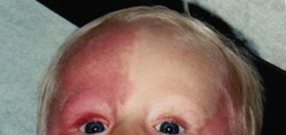
A 10-month-old infant was referred for evaluation of possible Sturge-Weber syndrome. According to his parents, the discoloration on the child’s face was present at birth. Physical examination revealed an otherwise healthy infant with extensive port-wine stains on his face. Ophthalmologic and neurologic examination findings were normal.
A 10-month-old infant was referred for evaluation of possible Sturge-Weber syndrome. According to his parents, the discoloration on the child’s face was present at birth (A).
Physical examination revealed an otherwise healthy infant with extensive port-wine stains on his face. Ophthalmologic and neurologic examination findings were normal. An x-ray film of the skull and cranial CT scan also were normal.
Alexander K. C. Leung, MD, and C. Pion Kao, MD, of Calgary, Alberta, tell us that the classic triad of Sturge-Weber syndrome includes:
•A port-wine stain over the face that involves mainly the cutaneous distribution of the ophthalmic division of the trigeminal nerve.
•Ipsilateral leptomeningeal angiomatosis and associated intracranial calcifications.
•Ocular abnormalities (glaucoma, buphthalmos, and choroidal vascular anomalies).
Although the port-wine stain in Sturge-Weber syndrome is usually unilateral, at times, it may be bilateral.
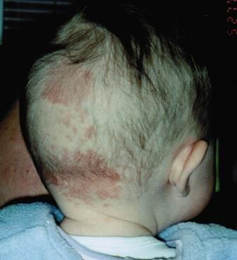
Port-wine stains, named for their purplish red color, are always present at birth. They consist of mature dilated dermal capillaries and represent a permanent developmental defect. The lesions are red to purple, macular, and sharply circumscribed. Although port-wine stains may occur anywhere on the body, they tend to favor the face. Unlike salmon patches (B), port-wine stains do not fade; in fact, they often darken and become pebbly in texture with age. Unlike capillary and cavernous hemangiomas, these lesions do not enlarge but remain stable and flat.
In addition to Sturge-Weber syndrome, port-wine stains may occur as a component of Klippel-Trnaunay-Weber syndrome, Cobb syndrome, Rubinstein-Taybi syndrome, Beckwith-Wiedemann syndrome, trisomy 13 syndrome, or Proteus syndrome.
Treatment with a flash lamp–pumped pulsed dye laser is highly effective and should be considered for children with port-wine stains if the lesion is bothersome or unsightly. This therapy specifically targets the lesion and avoids thermal injury to the surrounding normal tissue.


Recognize & Refer: Hemangiomas in pediatrics
July 17th 2019Contemporary Pediatrics sits down exclusively with Sheila Fallon Friedlander, MD, a professor dermatology and pediatrics, to discuss the one key condition for which she believes community pediatricians should be especially aware-hemangiomas.