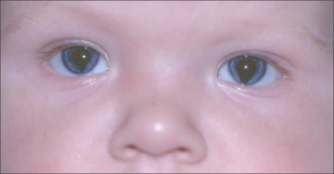
Bilateral Iris Colobomas
The patient's medical history was unremarkable. He had been born via normal vaginal delivery. His father had a history of strabismus. The child had fix-and-follow vision in both eyes. Extraocular muscle movement appeared normal. A Hirschberg test showed orthophoria. A Bruckner test showed symmetrical red reflexes in both eyes. Cycloplegic refraction revealed moderate hyperopic correction, which was symmetrical in both eyes. No retinal or optic nerve head defects were noted on dilated fundus examination.

FigureThis 5-month-old boy had been born with elongated pupils. His parents reported that the child's eyes appeared to work in unison (there was no eye crossing) and that he seemed to see well enough to grasp for objects. However, they were concerned about his vision.
The defect was more extensive in the left eye. Physical findings were otherwise normal.
The patient's medical history was unremarkable. He had been born via normal vaginal delivery. His father had a history of strabismus. The child had fix-and-follow vision in both eyes. Extraocular muscle movement appeared normal. A Hirschberg test showed orthophoria. A Bruckner test showed symmetrical red reflexes in both eyes. Cycloplegic refraction revealed moderate hyperopic correction, which was symmetrical in both eyes. No retinal or optic nerve head defects were noted on dilated fundus examination.
This child has coloboma of the irides. This common anomaly is typically located inferonasally. This anatomic site corresponds to the last area of closure of the embryonic fissure. The malformation resembles a keyhole. The lens, ciliary body, choroid, retina, and optic nerve may also be involved. Visual acuity is usually not affected unless a coloboma of the retina (macula) and optic nerve is also present. Large iris colobomas can cause photophobia. Cosmetic-tinted contact lenses may mask the defect and alleviate the photophobia.
Most cases of isolated colobomas are sporadic, and there is no family history of colobomas-as in this patient's case. This congenital defect may be transmitted as an autosomal dominant characteristic. Colobomas are also associated with several genetic syndromes, the most common of which is CHARGE syndrome. Affected children may have coloboma, heart defects, choanal atresia, retarded growth, genital anomalies, and ear anomalies, and deafness.1
Children with colobomas should undergo a dilated fundus examination. If this cannot be performed in the office or if the examination findings are abnormal, an ophthalmological referral is necessary.
This child was seen again at 11 months of age. He appeared healthy. No new eye or vision problems were reported, and ophthalmic findings remained unchanged. He will be followed with annual eye examinations.
References:
- Vissers LE, van Ravenswaaij CM, Admiraal R, et al. Mutations in a new member of the chromodomain gene family cause CHARGE syndrome. Nat Genet. 2004;36:955-957.