Branchial Cleft Cyst in an Infant
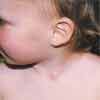
A 10-month-old girl with a mass on left side of neck. Swelling first noticed by mother when child was 7 months old. The infant is eating well, thriving, and otherwise healthy.

HISTORY
A 10-month-old girl with a mass on left side of neck. Swelling first noticed by mother when child was 7 months old. The infant is eating well, thriving, and otherwise healthy.
PHYSICAL EXAMINATION
A 4-mm cystic mass noted along the anterior border of the left sternocleidomastoid muscle. Mass is not red or tender, and is freely movable beneath the skin.
A branchial cleft cyst results from persistence of the cervical sinus of His.1,2
WHAT'S YOUR DIAGNOSIS?

CLINICAL MANIFESTATIONS
Branchial cleft anomalies may present as a cyst, sinus, fistula, or cartilaginous remnant.3 Approximately 80% of branchial cleft anomalies present as a cyst4 and about 95% are formed from the region of the second branchial arch.1,5 The remaining 5% arise from the regions of the first, third, or fourth arches.1,5
A branchial cleft cyst typically presents as a painless, mobile, and fluctuant mass located along the ante- rior border of the sternocleidomastoid muscle, usually just above the clavicle.4 Approximately 97% to 98% of the lesions are unilateral,6 and of these, 83% to 97% are on the left side--presumably consequentto asymmetrical vascular development.7
Although branchial cleft cysts are congenital and might be noted at birth, most are not detected until the first or second decade of life.8 Some are detected when they become more prominent in late childhood. Other cases become apparentduring intercurrent upper respiratory tract infections or when the cyst becomes infected.3
DIAGNOSIS AND DIFFERENTIAL DIAGNOSIS
The diagnosis is established by physical examination. Ultrasonography can help delineate the cystic nature of the lesion if the diagnosis is in doubt. The differential diagnosis includes cervical lymphadenopathy, fibrous dysplasia of the sternocleidomastoid muscle (fibromatosis coli), dermoid cyst, and cystic hygroma. A thyroglossal duct cyst is a midline structure, and should be easily differentiated.9
COMPLICATIONS
Secondary bacterial infection is a possible complication. Squamous cell carcinoma is a rare complicationreported in adulthood.10
HISTOPATHOLOGY
A branchial cleft cyst is lined by squamous or columnar epithelium.11,12 The cyst usually contains either a clear fluid or a toothpaste-like material, and may contain cholesterol crystals.
MANAGEMENT
Complete surgical excision with careful attention to identifying deeper components is the treatment of choice.3 Aspiration, or incision and drainage, is associated with an increased risk of recurrence and of such complications as wound infection or hemorrhage.7 Secondary infection requires systemic antibiotic therapy.
References:
REFERENCES:
1.
Cote DN, Gianoli GJ. Fourth branchial cleft cysts.
Otolaryngnol Head Neck Surg.
1996;114:95-97.
2.
Leung AK, Wong AL. Branchial cleft cyst.
Can J Diagn.
2004;21(2):46.
3.
Davenport M.ABC of general surgery in children: lumps and swellings of the head and neck.
BMJ.
1996;312:368-371.
4.
Agaton-Bonilla FC, Gay-Escoda C. Diagnosis and treatment of branchial cleft cysts and fistulae. A retrospective study of 183 patients.
Int J Oral Maxillofac Surg.
1996;25:449-452.
5.
Chaudhary N, Gupta A, Motwani G, Kumar S. Fistula of the fourth branchial pouch.
Am J Otolaryngol.
2003;24:250-252.
6.
Fritsch MH. Branchial cleft cyst with fistula.
Otolaryngol Head Neck Surg.
1992;107:133-134.
7.
Pereira KD, Losh GG, Oliver D, Poole MD. Management of anomalies of the third and fourth branchial pouches.
Int J Pediatr Otorhinolaryngol.
2004;68:43-50.
8.
Yilmaz I, Cakmak O, Ozgirgin N, et al. Complete fistula of the second branchial cleft: case report of catheter-aided total excision.
Int J Pediatr Otorhinolaryngol.
2004;68:1109-1113.
9.
Leung AK, Robson WL. Thyroglossal cyst.
Resident Staff Physician.
1992; 38(5):85.
10.
Khfif RA, Pricher R, Minkowitz S. Primary branchiogenic carcinoma.
Head Neck.
1992;12:943-960.
11.
Eichenfield L, Larrade M. Branchial clefts and auricular sinuses. In: Schachner LA, Hansen R, Happle R, eds.
Pediatric Dermatology.
New York: Mosby; 2003:222-224.
12.
Leung AK, Wong WL, Robson WL. Branchial cyst.
Resident Staff Physician.
1993;39(9):82.
13.
De Caluwé D, Hayes R, McDermott M, Corbally MT. Complex branchial fistula: a variant arch anomaly.
J Pediatr Surg.
2001;36:1087-1088.
14.
Turkington JR, Paterson A, Sweeney LE, Thornbury GD.Neck masses in children.
Br J Radiol.
2005;78:75-85.
15.
White SA, Narula AA. Multiple branchial cleft anomalies.
Eur J Surg.
1995; 161:443-445.
16.
Nicollas R, Guelfucci B, Roman S, Triglia JR. Congenital cysts and fistulas of the neck.
Int J Pediatr Otorhinolaryngol.
2000;55:117-124.
17.
Johnson IJ, Soames JV, Birchall JP. Fourth branchial arch fistula.
J Laryngol Otol.
1996;110:391-393.
18.
Edmonds JL, Girod DA, Woodroof JM, Bruegger DE. Third branchial anomalies. Avoiding recurrences.
Arch Otolaryngol Head Neck Surg.
1997; 123:438-441.
Recognize & Refer: Hemangiomas in pediatrics
July 17th 2019Contemporary Pediatrics sits down exclusively with Sheila Fallon Friedlander, MD, a professor dermatology and pediatrics, to discuss the one key condition for which she believes community pediatricians should be especially aware-hemangiomas.