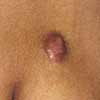
Photoclinic: Keloid at the Site of a Chickenpox Lesion
An 18-year-old girl presented with an asymptomatic nodule on the posterior aspect of the right upper arm. The lesion had developed a month after an episode of chickenpox at 11 years of age and had slowly enlarged. The lesion was 7 mm in diameter; it was firm, rubbery, reddish brown, and nontender.

An 18-year-old girl presented with an asymptomatic nodule on the posterior aspect of the right upper arm. The lesion had developed a month after an episode of chickenpox at 11 years of age and had slowly enlarged. The lesion was 7 mm in diameter; it was firm, rubbery, reddish brown, and nontender.
Alexander K. C. Leung, MD, and Wm. Lane M. Robson, MD, of Alberta Children's Hospital, Calgary, Alberta, write that this sharply demarcated nodule is a keloid at the site of a chickenpox lesion. Keloids result from proliferation of fibroblasts and deposition of collagen in the dermis after chickenpox infection, sternal acne, or cutaneous trauma (such as a laceration, surgical incision, ear piercing, deltoid vaccination, or burn and scald injuries).1 The condition may be familial and is more common in those with dark skin. Rarely, keloids are a feature of Rubinstein-Taybi syndrome, Ehlers-Danlos syndrome, Goeminne syndrome, or pachydermoperiostosis.
The surface of a keloid is typically smooth, and the lesion is firm. Although a keloid can occur anywhere on the body, the upper trunk, upper arms, lower legs, and ear lobes are frequently involved. The shape is often specific to the site: lobular in ear lobes, butterfly-shaped in the sternum, and vertical in the deltoid.
In contrast to a hypertrophic scar, which is asymptomatic, tends to stay within the margins of the original wound, and slowly regresses, a keloid extends well beyond the original wound, slowly enlarges, and can be pruritic or even painful.
A small keloid may not require treatment. A large or cosmetically unappealing keloid can be treated with intralesional injections of triamcinolone, either alone or in combination with surgical excision or pulsed dye laser therapy.
Intralesional injections of triamcinolone every 2 to 4 weeksfor 6 months resulted ina substantial reduction in the size of this patient's lesion.
References:
REFERENCE:
1.
Leung AK, Kao CP, Sauve RS. Scarring resulting from chickenpox.
Pediatr Dermatol.
2001;18:378-380.
Recognize & Refer: Hemangiomas in pediatrics
July 17th 2019Contemporary Pediatrics sits down exclusively with Sheila Fallon Friedlander, MD, a professor dermatology and pediatrics, to discuss the one key condition for which she believes community pediatricians should be especially aware-hemangiomas.