Case In Point: Infantile Hypertrophic Pyloric Stenosis
A 7-week-old white boy presented to the emergency department (ED) with vomiting and weight loss. His parents brought him to the ED 3 weeks earlier after he had vomited for several days. Possible milk protein allergy was diagnosed at that visit, and a change from cow milk formula to an elemental formula was recommended. Vomiting subsequently increased in frequency. Nonbilious but forceful vomiting occurred with each feeding. The patient lost nearly 2 lb during the 3 weeks that followed the first ED visit.

A 7-week-old white boy presented to the emergency department (ED) with vomiting and weight loss. His parents brought him to the ED 3 weeks earlier after he had vomited for several days. Possible milk protein allergy was diagnosed at that visit, and a change from cow milk formula to an elemental formula was recommended. Vomiting subsequently increased in frequency. Nonbilious but forceful vomiting occurred with each feeding. The patient lost nearly 2 lb during the 3 weeks that followed the first ED visit.
The patient was born at term by spontaneous vaginal delivery. The perinatal course had been unremarkable. Birth weight was 7 lb 10 oz. The baby weighed 9 lb 8 oz at 3 weeks and 8 lb at 7 weeks.
The patient was active and alert but had lost a significant amount of subcutaneous tissue. His abdomen was soft and nondistended. There were no masses in the upper quadrant and no organomegaly. Other physical examination findings were unremarkable.
The patient's white blood cell count was 12,400/µL, with 24% neutrophils and 74% lymphocytes. The hematocrit was 41.4%, and the platelet count was 685,000/µL. The serum sodium level was 129 mEq/L; serum potassium, 2.7 mEq/L; serum chloride, 72 mEq/L; serum bicarbonate, 37 mEq/L; blood urea nitrogen, 24 mg/dL; and serum creatinine, 0.5 mg/dL (44.2 µmol/L). Capillary blood gas pH was 7.56; partial pressure of alveolar carbon dioxide, 52.8 mm Hg; partial pressure of alveolar oxygen, 59.2 mm Hg; and bicarbonate level, 46.2 mEq/L.
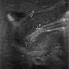
The persistent vomiting with hypochloremic metabolic alkalosis prompted the performance of abdominal ultrasonography. The test showed the length of the pyloric channel to be 19.5 mm and the muscle width itself to be more than 6.9 mm (Figure). These findings were diagnostic of pyloric stenosis. The patient's electrolyte abnormalities were corrected, and a pyloromyotomy was scheduled.
INFANTILE HYPERTROPHIC PYLORIC STENOSIS (IHPS)
IHPS classically presents with the gradual onset of nonbilious projectile vomiting at about the third week of life. However, 20% of infants are symptomatic from birth, and most are symptomatic within 2 months after birth.1 IHPS is the most common cause of nonbilious vomiting and metabolic alkalosis in infants.2-6 It is the most common reason for abdominal surgery in the first 6 months of life.
INCIDENCE
The incidence of IHPS varies from 1 in 200 to 1 in 750 live births. The disorder is 4 to 6 times more common in boys than in girls. The overall average is 3 in 1000 live births.1,2 Although believed to be more common in first-born boys, pyloric stenosis recently has been shown to be associated with smaller family size and higher socioeconomic class, rather than with birth order. The incidence is 2- to 3-fold higher in African Americans than in whites, and it is rare in Asians.1 The disorder has a familial tendency: 7% of children with this disorder have an affected parent. Among infants with IHPS, the incidence of recurrence is 10% in subsequent male siblings and 2% in subsequent female siblings.
ETIOLOGY/ PATHOPHYSIOLOGY
Multiple theories have been proposed to explain the development of pyloric stenosis. Postnatal infusion of gastrin produces an identical lesion in newborn puppies; it has therefore been suggested that hypergastrinemia may play an important etiologic role in IHPS.1,2 Similarly, prostaglandin E2 infusion--used to maintain a patent ductus arteriosus in certain cardiac anomalies--has been linked to a higher incidence of IHPS.1 Recent studies show a relative lack of nitric oxide synthase (a smooth muscle relaxant) innervation in the hypertrophied muscles.1 Recent studies suggest that erythromycin therapy may be associated with development of pyloric stenosis in infants younger than 30 days.3,4
Patients who have IHPS typically present with hypochloremic metabolic alkalosis. Although the serum potassium level may be normal or low, there often is total-body potassium depletion. The observed metabolic alkalosis is a result of 2 related but independent processes: loss of acid and retention of bicarbonate.
Initially, vomiting of gastric contents results in excessive loss of hydrogen chloride and potassium chloride, which leads to a metabolic alkalosis. Under normal circumstances, carbonic acid in the villi of the stomach dissociates into hydrogen and bicarbonate. The hydrogen ions cross the luminal membrane of the enterocyte and enter the stomach, from where they are transported to the duodenum. The entry of acid into the duodenum stimulates the secretion of an equal amount of pancreatic bicarbonate. This normal stimulus is absent in pyloric stenosis because of the mechanical obstruction: the diminished secretion of pancreatic bicarbonate into the GI tract contributes further to the metabolic alkalosis created by stomach acid lost through vomiting.
Moreover, as intravascular volume decreases with dehydration, the concentration of bicarbonate in the plasma increases, resulting in a "contraction alkalosis." The loss of potassium during vomiting also may contribute to the development of metabolic alkalosis. As the plasma potassium concentration falls, potassium moves out of the cells to replenish extracellular stores and electroneutrality is maintained by hydrogen moving into the cells. This shift causes paradoxical extracellular alkalosis with intracellular acidosis.
The kidneys also play a role in the metabolic alkalosis seen with pyloric stenosis. To maintain volume in the face of fluid losses from vomiting, the kidneys reabsorb bicarbonate in the distal tubules despite alkalosis. If the excess bicarbonate is excreted in the urine, it obligates sodium loss to maintain electroneutrality. Because water loss follows sodium excretion, further volume is lost. In addition, decreased chloride delivery to the macula densa of the kidneys results in renin release and secondary hyperaldosteronism. This, in turn, leads to increased distal hydrogen secretion and the paradoxical finding of acid urine in the presence of alkalemia. Finally, in response to hypokalemia, the distal tubules of the kidneys reabsorb potassium in exchange for hydrogen, resulting in further loss of acid.
CLINICAL FEATURES
Presentation. Nonbilious vomiting is the initial symptom of IHPS. The vomiting may or may not be projectile, but it is usually progressive and occurs during or shortly after each feeding. The full-terminfant usually has an avid appetite after vomiting. In contrast, the preterm infant usually does not display exaggerated peristalsis: although the child may appear hungry, he or she does not have the avid appetite seen in the full-term infant. The frequent preexistence of gastroesophageal reflux in the preterm infant may confound the diagnosis.
Physical examination. Typically, the infant is malnourished. He will have a palpable, firm, movable mass ("olive") in the right upper quadrant of the abdomen and visible intestinal peristalsis. To enhance the sensitivity of the physical examination, it may be helpful to empty the child's stomach with a nasogastric or orogastric tube and to palpate the abdomen while the child is held in a prone position. Comforting the infant to prevent crying also helps.
Diagnosis. When palpable, a pyloric mass is pathognomonic of hypertrophic pyloric stenosis. As noted, patients classically have a hypochloremic metabolic alkalosis, occasionally with severe deficits of potassium and sodium. Hyperbilirubinemia is present in about 5% of patients with pyloric stenosis.
Barium swallow shows a narrowed and elongated pyloric canal. Ultrasonography is the current diagnostic study of choice.7,8 Test results may be more accurate than clinical examination in detecting IHPS at an early stage, when the typical "olive" is not yet palpable and the peristaltic waves of the stomach are not yet visible.9 Ultrasonographic findings of a pyloric muscle thickness of more than 4 mm and a pyloric length of more than 14 mm have a diagnostic sensitivity and specificity of 95% and 100%, respectively.2,6
MANAGEMENT
Management of IHPS includes meticulous correction of fluids and electrolytes, followed by corrective surgery. Appropriate medical management reverses the volume contraction and metabolic derangements. All metabolic derangements--including hypokalemia and alkalosis, along with dehydration--must be corrected before surgical repair is undertaken.
Pyloromyotomy is curative for gastric outlet obstruction. The prognosis following surgery is excellent.
A REMINDER . . .
The diagnosis of pyloric stenosis is frequently delayed. Consider pyloric stenosis as a cause of emesis in infants. Remember that the presence of gastroesophageal reflux does not rule out pyloric stenosis. And keep in mind that even though hypochloremic metabolic alkalosis strongly suggests pyloric stenosis as the underlying problem, normal electrolyte levels do not exclude the diagnosis.5,10
References:
REFERENCES:
1.
Rudolph CD. Infantile hypertrophic pyloric stenosis. In: Rudolph CD, Rudolph AM, eds.
Rudolph's Pediatrics.
21st ed. New York: McGraw-Hill; 2002: 1402-1403.
2.
Wylie R. Pyloric stenosis and congenital anomalies of the stomach. In: Behrman RE, Kliegman RM, Jenson HB, eds.
Nelson Textbook of Pediatrics.
17th ed. Philadelphia: WB Saunders Co; 2004:1229-1231.
3.
Mahon BE, Rosenman MB, Kleiman MB. Maternal and infant use of erythromycin and other macrolide antibiotics as risk factors for infantile hypertrophic pyloric stenosis.
J Pediatr.
2001;139:380-384.
4.
Honein MA, Paulozzi LJ, Himelright IM, et al. Infantile hypertrophic pyloric stenosis after pertussis prophylaxis with erythromycin: a case review and cohort study.
Lancet.
1999;354:2101-2105.
5.
Broderick KB. Pyloric stenosis. In: Schaider J, Hayden SR, Wolfe R, et al, eds.
Rosen and Barkin's 5 Minute Emergency Medicine Consult.
2nd ed. Philadelphia: Lippincott Williams & Wilkins; 2003:926-927.
6.
Dinkevich E, Ozuah PO. Pyloric stenosis.
Pediatr Rev.
2000;21:249-250.
7.
Breaux CW Jr, Georgeson KE, Royal SA, Curnow AJ. Changing patterns in the diagnosis of hypertrophic pyloric stenosis.
Pediatrics.
1988;81: 213-217.
8.
Chen EA, Luks FI, Gilchrist BF, et al. Pyloric stenosis in the age of ultrasonography: fading skills, better patients?
J Pediatr Surg.
1996;31:829-830.
9.
Hernanz-Schulman M, Sells LL, Ambrosino MM, et al. Hypertrophic pyloric stenosis in the infant without a palpable olive: accuracy of sonographic diagnosis.
Radiology.
1994;193:771-776.
10.
Touloukian RJ, Higgins E. The spectrum of serum electrolytes in hypertrophic pyloric stenosis.
J Pediatr Surg.
1983;18:394-397.
Recognize & Refer: Hemangiomas in pediatrics
July 17th 2019Contemporary Pediatrics sits down exclusively with Sheila Fallon Friedlander, MD, a professor dermatology and pediatrics, to discuss the one key condition for which she believes community pediatricians should be especially aware-hemangiomas.