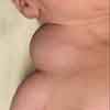
Cystic Hygroma in a Neonate
Patient delivered vaginally at term to a G2P1 24-year-old mother following an uncomplicated pregnancy. Apgar scores, 7 and 9 at 1 and 5 minutes, respectively. No history of maternal exposure to teratogens. Parents non-consanguineous. No family history of congenital or chromosomal abnormality.

HISTORY
Mass noted on the right side of newborn's neck.
Patient delivered vaginally at term to a G2P1 24-year-old mother following an uncomplicated pregnancy. Apgar scores, 7 and 9 at 1 and 5 minutes, respectively. No history of maternal exposure to teratogens. Parents non-consanguineous. No family history of congenital or chromosomal abnormality.
PHYSICAL EXAMINATION
Infant not in distress on examination. Soft cystic mass on neck measures 5 3 7 cm. Mass was brilliantly translucent and not attached to the overlying skin. Trachea in midline; no abnormality noted in the nasopharynx. A cystic hygroma is a developmental anomaly of the lymphatic system that is characterized by the formation of a multilocular cystic mass of variable size. The term "cystic hygroma" was first used by Wernher in 1843.1
WHATS YOUR DIAGNOSIS?
EMBRYOLOGY
The fetal lymphatic system develops around the fifth week of gestation as an endothelial outgrowth of the venous system.2,3 During embryonic development, 6 lymphatic sacs develop in close proximity to large veins.3 Lymphatic vessels extend in a centrifugal fashion from these lymphatic sacs through a process of branching.3 A cystic hygroma might result from either an abnormality in the control of lymphatic growth or from arrest in the normal development of the primitive lymphatic channels, whereby the peripheral lymphatic vessel becomes sequestrated and never connects with the remaining lymphatic system.4 The lesion arises in areas where tissuepressure is less and expansion of lymphatic tissue can occur.5
EPIDEMIOLOGY
The incidence is approximately 1 in 12,000 live births.4 The male to female ratio is equal.5 Cystic hygroma might follow maternal exposure to alcohol, trimethadone, or aminopterin.6 The condition is more common in patients with chromosomal abnormalities, such as Turner syndrome, Down syndrome, trisomy 13, trisomy 18, and Klinefelter syndrome.3,7 The incidence is alsoincreased in patients with Noonan syndrome, multiple pterygium syndrome, Roberts syndrome, Proteus syndrome, and Beckwith-Wiedemann syndrome.3,7 The condition might also be inherited as an autosomal recessive trait.7,8
CLINICAL MANIFESTATIONS
Cystic hygroma typically presents as a swelling that is compressible, feels softly cystic, fluctuates easily, and is brilliantly translucent.9 The lesion is not attached to the skin but might be attached to the underlying structures. Approximately 65% to 75% of cases are identified at birth, and 80% to 90% are diagnosed by the end of a child's third year of life.10 Cystic hygromas rarely manifest in adulthood.11
Approximately 75% of cystic hygromas occur in the neck--typically in the posterior cervical triangle.3,10 Cystic hygromas occur twice as often on the left side of the neck than on the right.4 Approximately 20% of the lesions occur in the axilla.3 Other less common sites include the mediastinum, trunk, extremities, abdomen, and retroperitoneum.3
The growth of a cystic hygroma is usually proportional to the growth of the child. The growth might increase during pregnancy.10 The lesion rarely regresses spontaneously.12
COMPLICATIONS
Although cystic hygroma is a benign lesion, it has the potential for extension or infiltration into the surrounding structures.13 Depending on the structures involved, airway obstruction, dysphagia, and feeding problems might result. Other potentialcomplications include infection and hemorrhage.
DIAGNOSIS AND DIFFERENTIAL DIAGNOSIS
Cystic hygroma might be first noted during prenatal ultrasonography. After birth, the diagnosis is established by physical examination. When the lesion is in the neck, the differential diagnosis includes branchial cleft cyst, dermoid cyst, thyroglossal duct cyst, lipoma, hemangioma, fibrous dysplasia of the sternocleidomastoid muscle (fibromatosis colli), cervical lymphadenopathy, and neuroblastoma.
HISTOPATHOLOGY
The lesion consists of multiloculated cysts that are lined by endothelial cells and that contain serous lymphatic fluid.3,14 Some cysts communicate with each other, whereas others are septated.14
DIAGNOSTIC STUDIES
For superficial lesions, ultrasonography adequately defines the size and extension of the lesion. For more complex lesions, CT or MRI imaging is necessary to define the relationship of the lesion with the adjacent structures. These studies are necessary before surgery is contemplated.4
TREATMENT
Indications for treatment include symptomatic lesions andcosmetic concerns. Surgery is the treatment of choice. If the lesion cannot be completely resected, recurrence is usually inevitable.4 Other treatment modalities include aspiration and injection of sclerosing agents such as OK-432.15 OK-432 is a lyophilized biologic preparation that contains the cells of Streptococcus pyogenes (group A, type 3) Su strain, which has been incubated with benzylpenicillin.14 OK-432 causes less fibrosis of the subcutaneous tissue and the overlying skin, and as such, the cosmetic result is better.3
References:
REFERENCES:
1.
Wernher A. Die angeborenen Kysten-hygrome und die ihnen verwandten.In:
Geschwulste in Anatomischer, Diagnosticher
und Therapeutischer Beziehung.
Giessen, Germany: GF Heyer, Vater; 1843:76-91.
2.
Fisher R, Partington A, Dykes E. Cystic hygroma: comparison between prenatal and postnatal diagnosis.
J Pediatr Surg.
1996;31:473-476.
3.
Gallagher PG, Mahoney MJ, Gosche JR. Cystic hygroma in the fetus and newborn.
Semin Perinatol.
1999;23:341-356.
4.
Ozen IO, Moralioglu S, Karabulut R, et al. Surgical treatment of cervicofacial cystic hygromas in children.
ORL J Otorhinolaryngol Relat Spec.
2005;67:331-334.
5.
Kennedy TL, Whitaker M, Pellitteri P, Wood WE. Cystic hygroma/lymphangioma: a rational approach to management.
Laryngoscope
. 2001;111:1929-1937.
6.
Edwards MJ, Graham JM Jr. Posterior nuchal cystic hygroma.
Clin Perinatol
. 1990;17:611-640.
7.
Langer JC, Fitzgerald PG, Desa D, et al. Cervical cystic hygroma in the fetus: clinical spectrum and outcome.
J Pediatr Surg
. 1990;25:58-61.
8.
Dallapiccola B, Zelante L, Perla G, Villani G. Prenatal diagnosis of recurrence of cystic hygroma with normal chromosomes.
Prenat Diagn
. 1984;4:383-386.
9.
Leung AK, Wong BE, Chan PY. Cystic hygroma.
Resident Staff Physician
. 1997;43:40.
10.
Avitia S, Osborne RF. Cystic hygroma exacerbated by pregnancy.
Ear Nose Throat J
. 2005;84:78-79.
11.
Aneeshkumar MK, Kale S, Kabbani M, David VC. Cystic lymphangioma in adults: can trauma be the trigger?
Eur Arch Otorhinolaryngol
. 2005;262:335-337.
12.
Emery PJ, Bailey CM, Evans JN. Cystic hygroma of the head and neck. A review of 37 cases.
J Laryngol Otol
. 1984;98:613-619.
13.
Ameh EA, Nmadu PT. Cervical cystic hygroma: pre-, intra-, and post-operative morbidity and mortality in Zaria, Nigeria.
Pediatr Surg Int
. 2001;17:342-343.
14.
Charabi B, Bretlau P, Bille M, Holmelund M. Cystic hygroma of the head and neck--a long-term follow-up of 44 cases.
Acta Otolaryngol Suppl
. 2000;543: 248-250.
15.
Karkos PD, Spencer MG, Lee M, Hamid BN. Cervical cystic hygroma/lymphangioma: an acquired idiopathic late presentation.
J Laryngol Otol
. 2005;119:561-563.
Recognize & Refer: Hemangiomas in pediatrics
July 17th 2019Contemporary Pediatrics sits down exclusively with Sheila Fallon Friedlander, MD, a professor dermatology and pediatrics, to discuss the one key condition for which she believes community pediatricians should be especially aware-hemangiomas.