Tinea Capitis and Tinea Corporis
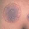
A 16-year-old boy complains of an itchy scalp and areas of hair loss. Physical examination reveals a 2- to 3-cm patch of alopecia studded with black dots, as well as posterior cervical and occipital lymphadenopathy.
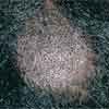
Case 1:
A 16-year-old boy complains of an itchy scalp and areas of hair loss. Physical examination reveals a 2- to 3-cm patch of alopecia studded with black dots, as well as posterior cervical and occipital lymphadenopathy.
What does this look like to you?
Case 1: This patient has tinea capitis, a dermatophyte infection of the scalp that in immunocompetent persons is usually caused by Trichophyton tonsurans. Clinical appearance may vary widely, but a common presentation is the gray type, which consists of localized patchy or diffuse scale accompanied by areas of alopecia. Regional lymphadenopathy may be present but is not specific. Another common presentation is "black dot" tinea capitis, which manifests as alopecic areas studded with black dots. The dots represent the residua of broken hairs, a result of fungal invasion of the hair shaft that leads to increased hair fragility and breakage at the surface of the scalp.
The diagnosis of tinea capitis is made by potassium hydroxide (KOH) evaluation and fungal culture.
Successful treatment requires a systemic antifungal agent. The "gold standard" agent has been oral griseofulvin. However, newer systemic antifungal agents, such as terbinafine and itraconazole, are as effective as griseofulvin and require shorter treatment times. Depending on the severity of the infection and the amount of associated inflammation, adjunctive therapy may be required. Some systemic antifungal medications, such as terbinafine and itraconazole, require laboratory monitoring because of potential toxicity. It is imperative to document infection by KOH evaluation or culture before systemic therapy is started.
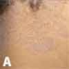
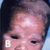
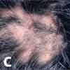
An antifungal shampoo, such as ketoconazole or selenium sulfide, should be used in conjunction with the systemic antifungal agent to help reduce fungal colony counts in the scalp. Antifungal shampoos should also be used prophylactically by close contacts and family members. Treatment is continued until scalp lesions resolve and culture results are negative.
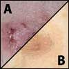
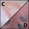
The differential diagnosis of tinea capitis includes seborrheic dermatitis (A), infantile seborrheic dermatitis (B), and trichotillomania (C). Seborrheic dermatitis is a chronic inflammatory condition characterized by erythematous patches and plaques with an overlying greasy yellowish scale. It is commonly localized to areas with high concentrations of sebaceous glands, such as the scalp and face. It does not commonly cause alopecia.
Trichotillomania, which is most commonly observed in adolescent girls, results in significant hair loss. This condition is categorized within the spectrum of obsessive-compulsive disorders. Treatment involves a multidisciplinary approach, including psychotherapy and adjuvant medications.
Case 2:
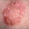
A 17-year-old boy presents with a 1-month history of erythematous, pruritic, scaly round lesions on his arms and chest. The rash began as a single affected area on the forearm; additional lesions have appeared during the past few weeks.
The patient is a member of the high school wrestling team. He competes against different opponents every week and is not aware of anyone else with similar symptoms.
Can you identify this outbreak?

Case 2: This patient has tinea corporis, which is characterized by erythematous patches that expand into annular plaques with central clearing and elevated borders distributed over the extremities and trunk. In immunocompetent persons, the most common cause of tinea corporis is Trichophyton rubrum. Athletes who engage in close contact sports, such as wrestling, are at high risk for acquisition of the infection from other players. Moreover, contaminated fomites (such as gym mats, headgear, and other personal equipment) can transmit infection.
A potassium hydroxide (KOH) evaluation is the most appropriate diagnostic test for tinea corporis. (The KOH dissolves cutaneous epithelial cells and allows for microscopic visualization of branching hyphae.) The diagnosis can be confirmed with a fungal culture.
Initial treatment consists of twice-daily application of a topical azole antifungal, such as ketoconazole or clotrimazole; an allylamine, such as terbinafine or naftifine; or ciclopirox olamine. The medication should be used for at least 2 weeks; each application should cover the lesion as well as at least a 2-cm perimeter of normal skin.
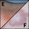
The differential diagnosis of tinea corporis includes other scaly, pruritic conditions and annular lesions such as nummular eczema (A), tinea versicolor (B), pityriasis rosea (C), psoriasis (D), allergic contact dermatitis (E), and granuloma annulare (F).
The conditions in the differential may be distinguished clinically by history and distribution, negative results on KOH evaluation and, if necessary, biopsy. The exception is tinea versicolor, which demonstrates hyphae and spores on KOH evaluation. Direct skin contact with an allergen (such as poison ivy, metals, fragrances, or various chemicals) can trigger the development of erythema, edema, vesicles, and pruritus that erupt in the distribution of contact. These lesions distinguish allergic contact dermatitis, a delayed hypersensitivy reaction, from fungal infection, which rarely presents with vesicles and papules at the advancing border.
Recognize & Refer: Hemangiomas in pediatrics
July 17th 2019Contemporary Pediatrics sits down exclusively with Sheila Fallon Friedlander, MD, a professor dermatology and pediatrics, to discuss the one key condition for which she believes community pediatricians should be especially aware-hemangiomas.