Nevus of Ota
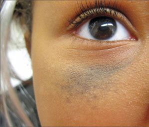
A 7-year-old African American girl presented with an asymptomatic gray lesion beneath her right eye that had appeared before her first birthday. She was a healthy child with macular gray discoloration on the right infraorbital skin. There was no abnormal pigment noted on the conjunctiva.

A 7-year-old African American girl presented with an asymptomatic gray lesion beneath her right eye that had appeared before her first birthday. She was a healthy child with macular gray discoloration on the right infraorbital skin. There was no abnormal pigment noted on the conjunctiva.
Nevus of Ota, also called oculodermal melanosis, is most commonly seen in Asians and African Americans, with 80% of cases occurring in females. It presents most often as unilateral bluish gray macules up to several centimeters in diameter in the distribution of the ophthalmic and maxillary branches of the trigeminal nerve. The lesion can involve the sclera and tympanum and, rarely, the cornea, retina, optic nerve, nasal mucosa, and oropharynx. Visual acuity is usually not affected. In up to half of cases, the lesions are congenital or appear within the first year of life; however, there is a second spike in incidence during adolescence. The lesion may slowly enlarge but typically remains stable during adulthood.
Pathological analysis shows an increased number of pigment-producing melanocytes in the dermis. There are rare reports of melanoma developing in nevi of Ota, so close observation is recommended.1 Laser surgery, specifically using pulsed Q-switched ruby, alexandrite, or Nd:YAG lasers, is currently the treatment of choice; multiple treatment sessions are often required. Q-switched lasers provide short pulses and high energy with selective photothermolysis of the pigment cells and minimal damage to the surrounding tissues.2
References:
REFERENCES:
1.
Bolognia JL, Jorizzo JL, Rapini RP, eds.
Dermatology
. Amsterdam: Elsevier; 2008:1720-1722.
2.
Chan H, Ying SY, Ho WS, et al. An in vivo trial comparing the clinical efficacy and complications of Q-switched 755 nm alexandrite and Q-switched 1064 nm Nd:YAG lasers in the treatment of nevus of Ota.
Dermatol Surg
. 2000;26:919-922.
Recognize & Refer: Hemangiomas in pediatrics
July 17th 2019Contemporary Pediatrics sits down exclusively with Sheila Fallon Friedlander, MD, a professor dermatology and pediatrics, to discuss the one key condition for which she believes community pediatricians should be especially aware-hemangiomas.