Persistent Ankle Mass in an Otherwise Healthy 9-Year-Old Girl
An apparently healthy 9-year-old girl noted to have left ankle mass during well-child checkup. Her last well-child visit was 3 years earlier. Medical history unremarkable. She denied fevers, weight loss, night sweats, and chills. No family history of bone deformities or growth disturbances.
HISTORY
An apparently healthy 9-year-old girl noted to have left ankle mass during well-child checkup. Her last well-child visit was 3 years earlier. Medical history unremarkable. She denied fevers, weight loss, night sweats, and chills. No family history of bone deformities or growth disturbances.
PHYSICAL EXAMINATION
Hard, fixed, irregular mass about 6 x 9 cm on the lateral aspect of her left distal tibia, nontender on palpation. Examination findings otherwise normal. On further questioning, the child reported that her ankle had been swollen for the past few months. She denied any trauma to the area.
LABORATORY RESULTS
Complete blood cell count, erythrocyte sedimentation rate, and C-reactive protein level normal.
IMAGING
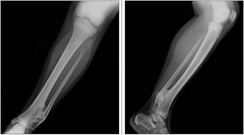
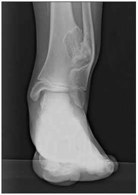
Radiographs of the left lower leg shown.

WHAT’S YOUR DIAGNOSIS? (Answer on next page)

ANSWER: OSTEOCHONDROMAS
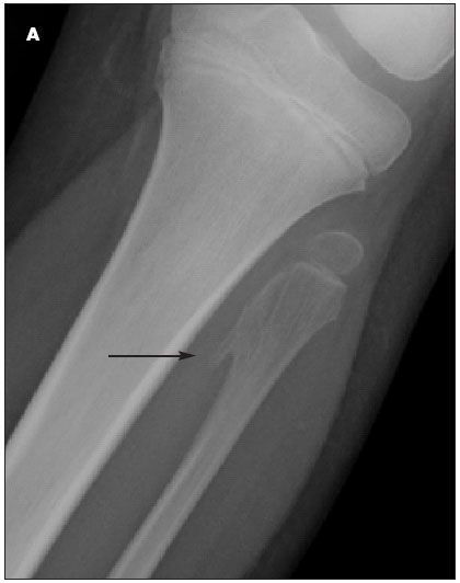
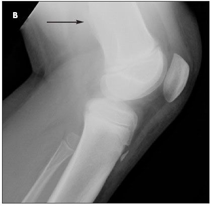
The radiographs revealed multiple bony excrescences with well-defined margins. These were identified as 1 large osteochondroma of the left lateral distal tibia that crossed the interosseous space, resulting in erosion and bowing of the fibula, and 2 smaller osteochondromas: 1 in the proximal fibular metaphysis (A) and 1 in the distal lateral femur (B).
INCIDENCE AND COMPLICATIONS
Osteochondroma is the most common benign bone tumor in children; it may arise spontaneously or after trauma to the osseous surface of any bone that forms from cartilage. Osteochondromas account for 20% to 50% of benign bone tumors and 10% to 15% of all bone tumors. 1 The tumor contains both medullary and cortical bone and has an overlying hyaline cartilage cap. The long bones of the lower extremities are most commonly affected.1,2 Osteochondromas of the long bones are more likely to involve the metaphysis than the diaphysis.1 The lesions usually stop growing once bone growth and maturation are completed.

Osteochondromas are often painless and may be found only after they cause significant physical deformity- or they may be identified incidentally on radiographs obtained for other reasons. However, they may cause irritation of the surrounding muscles, nerves, and tendons, which subsequently causes pain. Pedunculated osteochondromas can cause sudden pain if the tumor breaks off the stalk. Other complications include bony deformity, vascular compromise, bursa formation, nerve involvement, and malignant transformation.1-3 Osteochondromas may affect the growth plate and hinder bone development, resulting in asymmetrical growth or bowing.1 When the growth plate is affected at the ankle or knee, the leg length discrepancy is usually around 4 to 5 cm.4
Osteochondromas may be single or multiple. Multiple osteochondromas are more likely to be inherited. Hereditary multiple exostoses (HME) has an autosomal dominant inheritance pattern. About two-thirds of persons with multiple osteochondromas have a family history of HME. HME also commonly involves bone deformities.1
RISK OF MALIGNANT TRANSFORMATION
Solitary osteochondromas have a 1% chance of becoming malignant.1 The rate of malignant transformation in patients with HME is 3% to 5%.1 The likelihood of malignant transformation is increased in the following settings, which warrant closer follow-up:
- Osteochondromas that continue to grow after a child’s growth and bone maturation are complete.
- Osteochondromas that recur after resection.
- Osteochondromas that have a hyaline cartilage cap thicker than 3 cm at any time or greater than 1.5 cm after skeletal maturity.3
- Centrally located osteochondromas-those affecting the shoulders, pelvis, or hips.
- Osteochondromas that cause persistent pain.
The most common malignancy associated with multiple osteochondromas is a chondrosarcoma.1 Malignant transformation rarely occurs in patients younger than 20 years.1
DIFFERENTIAL DIAGNOSIS
Solitary osteochondromas can be confused with osteosarcoma, myositis ossificans, and a singular chondrosarcoma. Multiple osteochondromas should be distinguished from benign parosteal osteochondromatous proliferation, Ollier disease (enchondromatosis), and a reactive process.2,5,6 These entities often have other signs and symptoms as well as a different radiographic appearance. For instance, Ollier disease can be distinguished from HME based on location: the enchondromas in Ollier disease are located in the center of the bone, whereas osteochondromas are usually located near the surface of the bone.
Multiple exostoses can be part of other inherited conditions, such as metachondromatosis, Langer-Giedion syndrome, and Potocki-Shaffer syndrome.5,7 The exostoses in metachondromatosis are usually located in the hands and feet and often resolve spontaneously. They can also be distinguished from HME radiographically; they usually point toward the epiphysis and may be completely cartilaginous or cartilaginous with calcifications, thus not always radiopaque.7 Both Langer-Giedion syndrome and Potocki-Shaffer syndrome are associated with craniofacial abnormalities and mental retardation.5

DIAGNOSIS
The radiographic appearance of osteochondromas is often pathognomonic; radiographs typically show an outgrowth of cortical and medullary bone continuous with the underlying bone.1,2 Invariably, the lesion appears smaller radiographically than what is suggested by palpation because the cartilage cap covering the lesion is not visible on plain radiographs. In cases in which the diagnosis cannot be made from radiographs alone, MRI can be useful in the evaluation of a suspected osteochondroma because it can show the cartilage cap and nerve involvement.1,3 CT and ultrasonography may also be used in some circumstances.1
MANAGEMENT
Solitary osteochondromas are usually monitored clinically and may be followed radiographically. Those that cause pain or other complications may be surgically removed. The recurrence rate after removal of a solitary tumor is low (between 1% and 2%) and rarely reported. 8 Possible cosmetic interventions include debulking or chipping with an osteotome; however, these are done after the bone has matured to avoid damaging the growth plate.5 Although surgical excision is unnecessary for benign multiple osteochondromas, closer monitoring is warranted because of the increased risk of malignancy.
This patient’s condition falls within the spectrum of HME. She was referred to an orthopedic surgeon for regular clinical and radiographic follow-up for progression, complications, and possible malignant transformation. Although she is currently asymptomatic, she may require treatment in the future. Any type of cosmesis would probably involve more than 1 surgery to correct the ankle deformity and subsequent limb shortening caused by the damaged growth plate.
References:
REFERENCES:1. Murphey MD, Choi JJ, Kransdorf MJ, et al. Imaging of osteochondroma: variants and complications with radiologic-pathologic correlation. Radiographics. 2000;20:1407-1434.
2. Fuselier CO, Binning T, Kushner D, et al. Solitary osteochondroma of the foot: an in-depth study with case reports. J Foot Surg. 1984;23:3-24.
3. Quirini GE, Meyer JR, Herman M, Russell EJ. Osteochondroma of the thoracic spine: an unusual cause of spinal cord compression. AJNR. 1996;17:961-964.
4. Alvarez CM, De Vera MA, Heslip TR, Casey B. Evaluation of the anatomic burden of patients with hereditary multiple exostoses. Clin Orthop Relat Res. 2007;462:73-79.
5. Pannier S, Legeai-Mallet L. Hereditary multiple exostoses and enchondromatosis. Best Pract Res Clin Rheumatol. 2008;22:45-54.
6. Oviedo A, Simmons T, Benya E, Gonzalez-Crussi F. Bizarre parosteal osteochondromatous proliferation: case report and review of the literature. Pediatr Dev Pathol. 2001;4:496-500.
7. Kennedy LA. Metachondromatosis. Radiology. 1983;148:117-118.
8. Morton KS. On the question of recurrence of osteochondroma. J Bone Joint Surg Br. 1964;46:723-725.
Recognize & Refer: Hemangiomas in pediatrics
July 17th 2019Contemporary Pediatrics sits down exclusively with Sheila Fallon Friedlander, MD, a professor dermatology and pediatrics, to discuss the one key condition for which she believes community pediatricians should be especially aware-hemangiomas.


