Isolated Macrodactyly
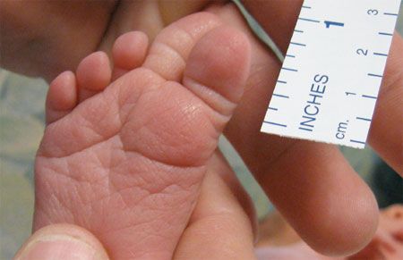
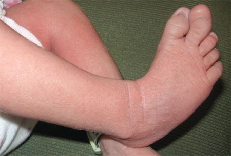
This well-formed, enlarged second digit on the right foot of a 2-day-old girl was noted at birth. All other digits were of normal size. The distal portion of the digit is deviated medially with a diminished triangular matrix unguis. No other musculoskeletal anomalies are apparent. There is no family history of congenital malformations or deformations.
This well-formed, enlarged second digit on the right foot of a 2-day-old girl was noted at birth. All other digits were of normal size. The distal portion of the digit is deviated medially with a diminished triangular matrix unguis. No other musculoskeletal anomalies are apparent. There is no family history of congenital malformations or deformations.
Macrodactyly is a rare congenital abnormality characterized by the enlargement of one or more (but not all) of the fingers or toes in the absence of other lesions. This structural abnormality is more common in the hands than in the feet and affects multiple digits more often than an isolated digit. There is a slight male predominance, with a ratio of 3:2 in some studies. The cause is not fully understood but appears to be a combination of local, genetic, and environmental factors.

There are 2 forms of macrodactyly, which are differentiated by the relative growth rate of the digit. The static form-macrodactyly simplex congenita (MSC)-is present at birth; the growth rate of the affected digit is similar to that of the other digits. The progressive form-macrodystrophia lipomatosa progressiva (MLP)-may or may not be present at birth; it is characterized by a disproportionate, accelerated growth rate of the affected finger or toe. Bone and nerve tissue may be involved in MSC, whereas MLP is usually the result of fibrofatty tissue proliferation and may present with progressive changes to the proximal structures.

Macrodactyly may be difficult to definitively diagnose at birth; however, the diagnosis may be confirmed by age 6 months after careful observation of digital growth. Ultrasonography and radiography of the affected digits may be beneficial in this regard.
Surgical options include debulking, ray resection, and toe amputation (not performed on the first digit). The goal of surgery is to produce a painless, cosmetically acceptable digit that regular footwear can accommodate. The digit can also be left untreated until the child’s bone growth reaches maturity. The treatment approach depends on the parents’ perception of how it will affect their child, psychologically as well as physiologically.