Persistent foot and leg swelling in a 17-year-old female
A 17-year-old girl presents with a 2-year history of unilateral swelling of the left lower extremity as well as a poorly healed ankle sprain of the affected extremity 3 years prior that slowly resolved but left persistent swelling. What's the diagnosis?
The case
A 17-year-old girl presents with a 2-year history of unilateral swelling of the left lower extremity. The patient reports a poorly healed ankle sprain of the affected extremity 3 years prior that slowly resolved but left persistent swelling. She states that the swelling fluctuates, reaching as high as the midcalf, but has persistent baseline enlargement compared with the opposite foot.
History
Medical history includes hypothyroidism, successfully managed with levothyroxine, which was discontinued 3 years earlier as thyroid function tests normalized. She reached thelarche at age 11 years and menarche at age 13 years. Developmental and family history are unremarkable and noncontributory.
Physical exam
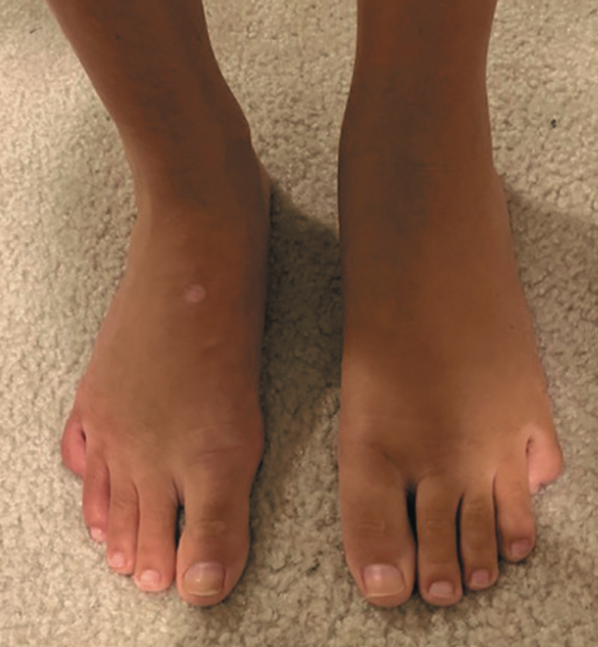
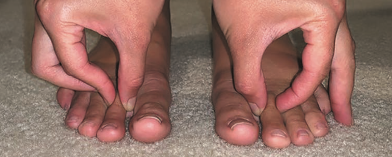
Upon examination, vital signs and growth parameters are normal. The left foot is notable for nonpitting edema to the level of the malleoli (Figure 1); it is nontender and without discoloration. Kaposi-Stemmer sign (inability to pinch the skin at the base of the second toe) is positive (Figure 2). The remainder of her examination is normal with no evidence of thyromegaly, lymphadenopathy, hepatosplenomegaly, or swelling of the other extremities.
Figure 1

Laboratory testing and imaging
Thyroid function tests, complete blood cell count, and chemistry panel obtained at presentation are normal. Abdominal and pelvic ultrasounds do not reveal any evidence of an obstructing mass or other abnormalities. Magnetic resonance imaging without contrast is negative other than an incidental finding of tarsal coalition, mostly fibrous, between the calcaneus and talus.
Differential diagnosis
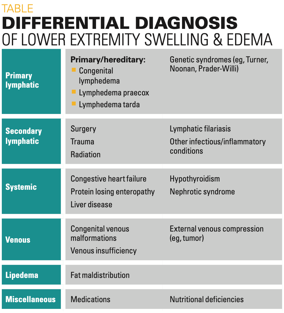
The differential diagnosis for chronic unilateral lower extremity swelling in an adolescent includes lymphatic and venous etiologies of edema, as well as lipedema (Table). Lymphatic etiologies include primary and hereditary processes (often distinguished based on age of presentation), secondary causes, and associated genetic syndromes. Venous etiologies include congenital venous malformations, venous insufficiency, and external venous compression from tumor, trauma, or other mechanical obstruction.1 Edema from congenital venous malformation would likely present earlier in life, and venous insufficiency would be uncommon in an otherwise healthy adolescent. Furthermore, venous edema is usually pitting and often accompanied by hyperpigmentation of the extremity because of hemosiderin deposition.2 Although lipedema is caused by fat maldistribution rather than edema, it can present similarly to venous or lymphatic edema. It is often exquisitely tender and nonpitting and spares the foot.3 Systemic causes of edema such as congestive heart failure, nephrotic syndrome, protein-losing enteropathies, and cirrhosis are more likely to cause bilateral, pitting edema.2 Myxedema can also be noted in advanced stages of hypothyroidism but is typically ac- companied by other findings (eg, bradycardia, weight gain).2 Certain medications—notably, calcium channel blockers, corticosteroids, and chronic use of nonsteroidal anti-inflammatory drugs—can cause lower extremity edema.3 Lymphedema can be differentiated from nonlymphatic causes of edema by the presence of the Kaposi-Stemmer sign (Figure 2), as inflammation and adipose deposition from the lymphedema can lead to thickening of the skin.1
Table

A thorough evaluation ruled out systemic, malignant, or compressive causes of the unilateral swelling. Based on the exam findings and the patient’s history, including age of onset and preceding injury to the affected extremity, as well as the positive Kaposi-Stemmer sign, the patient received a clinical diagnosis of lymphedema praecox.
Discussion
Lymphedema praecox is the most common type of primary lymphedema. It develops before age 35 years and often shortly after puberty. It classically affects the lower extremities and is typically unilateral; however, it can affect the upper extremity or present bilaterally. Edema is usually pitting on initial examination but often becomes nonpitting over time because of fibrosis causing the skin to turn leathery and indurated, leading to the nonpitting edema that represents chronic, irreversible stages of the disease.3
Figure 2

The condition affects female patients more often than male patients by a 4:1 ratio. This disparity is especially prevalent in children and young adults aged between 10 and 20 years, which led to the belief that estrogen is implicated in the disease pathogenesis.
This is further supported by evidence of children with Turner syndrome who during infancy had congenital lymphedema that spontaneously subsided and then returned shortly after menarche.5
Lymphedema praecox is thought to be associated with a variety of genetic loci, but a causal relationship between the disease and any specific genetic defect has yet to be defined, and most cases are sporadic rather than hereditary.4 Meige disease refers to a familial subtype of lymphedema praecox and is also known as hereditary lymphedema type 2. The late onset of lymphedema praecox despite its congenital nature is thought to result from a secondary event later in life (eg, injury or infection) that leads to a sudden worsening of a previously subclinical lymphatic defect.6
The differential diagnosis for lymphedema praecox includes other forms of both primary and secondary lymphedema. The 3 types of primary lymphedema are distinguished by age of onset and include congenital lymphedema, lymphedema praecox, and lymphedema tarda.3,4 All 3 conditions are caused by primary lymphatic abnormalities that may not manifest until later in life and mainly involve the lower extremity.
Congenital lymphedema is defined as primary lymphedema that presents within the first year of life. Milroy disease is a specific subtype of congenital lymphedema and is also called hereditary lymphedema type 1A. Up-slanting toenails and enlarged leg veins have also been associated with Milroy disease as has hydroceles in male patients. In most cases, the swelling is bilateral and remains constant in severity throughout the patient’s lifetime.4 Both Milroy disease and Meige disease are inherited in an autosomal dominant manner.4 Lymphedema tarda is defined as primary lymphedema presenting after age 35 years and is associated with impaired wound healing due to stasis of lymphatic flow and ulcers that fail to heal because of regional oxygen deficiency in the diseased tissue.7
Secondary lymphedema is caused by impaired lymphatic flow from an acquired cause. It is far more common than primary lymphedema and usually becomes symptomatic later in life compared with primary lymphedema. Most cases of secondary lymphedema result from surgery or local radiation causing mechanical lymphatic obstruction. The most common cause worldwide is lymphatic filariasis, also known as elephantiasis, a mosquito-borne infection caused by the roundworm Wuchereria bancrofti. The infection is endemic to India and sub-Saharan Africa.3 Other causes include an obstructing mass, trauma, polysplenia syndrome, Hodgkin lymphoma, rosacea, chronic venous hypertension, venous ulcers, fluid overload, Kaposi sarcoma, obesity, rheumatoid or psoriatic arthritis, allergic contact dermatitis, and drug induced lymphedema.3
Diagnosis and management
Several radiological modalities are used for the evaluation and diagnosis of lymphedema. Direct lymphangiography allows for real-time visualization of lymphatic flow and assesses the patency of lymphatic channels. It was once the method of choice for imaging lymphedema but is rarely used now because of potential adverse effects, including hypersensitivity reactions and patient discomfort.8 Direct lymphangiography has been replaced by less invasive procedures such as magnetic resonance lymphangiography and fluorescence microlymphography, a technique that allows for lymphatic visualization using an injection of fluorescein isothiocyanate-dextran.8,9 Lymphoscintigraphy, a radionucleotide imaging study, can be used to evaluate lymphatic anatomy, vessel patency, dynamics of flow, and flow reversal. It is considered the most definitive test for diagnosing lymphedema and has a specificity of 100%.10
Although there is no definitive cure for primary or secondary lymphedema, effective treatment and symptom relief can be achieved by either surgical or nonsurgical treatment modalities. Symptomatic management of early-stage lymphedema can be achieved with nonsurgical decongestive therapy, which includes massage, lymph drainage, compression therapy, and advanced pneumatic compression pumps.12 Compression garments should be used continuously throughout the day and may be removed at night when the extremity can be elevated. Pneumatic pump compression should be done prior to fibrosclerotic evolution to prevent its development. Both medical and pneumatic compression therapy may be contraindicated in patients with congestive heart failure, deep vein thrombosis, or active infection.13 Low-level laser therapy has also been shown to decrease pain and physical parameters of lymphedema. It is thought to increase lymphatic drainage as well as stimulate the formation of new lymph vessels.12
Surgical intervention has been effective in treating lymphedema not responsive to less invasive modalities. Options include direct excision and liposuction. Physiologic techniques, such as lymphatic venous anastomosis or bypass, have also been shown to be effective.12
Prognosis and complications
Prognosis for patients with lymphedema varies based on disease chronicity, etiology, resulting complications, and the patient’s adherence with maintenance modalities. Primary lymphedema usually does not progress and often stabilizes after several years. Primary lymphedema is associated with lower morbidity and better outcomes than secondary lymphedema.
Potential complications of long-standing or untreated lymphedema include recurrent cellulitis and other recurrent bacterial and fungal infections. Patients with a history of chronic lymphedema lasting longer than 10 years have a 10% risk of developing angiosarcoma.3 Other malignancies associated with long-standing lymphedema include basal and squamous cell carcinoma, Kaposi sarcoma, melanoma, and cutaneous lymphomas. Additional complications can include recurrent skin ulcers and elephantiasis nostra verrucosa, as well as functional impairment, activity restrictions, and cosmetic deformity, which can have significant psychosocial repercussions.
Patient course
Diagnostic imaging was recommended, but the patient’s parents declined because of concern of invasive techniques and if any such procedure would provide additional diagnostic information, given the diagnosis of lymphedema praecox. The patient was instructed to apply Jobst compression garments to the affected extremity to help eliminate the remaining swelling. Physical therapy and lymphatic massage were also recommended. The leg swelling decreased dramatically with therapy but intermittently recurs when therapy is discontinued.
References
- Green AK, Goss JA. Diagnosis and staging of lymphedema. Semin Plast Surg. 2018;32(1):12-16. doi:10.1055/s-0038-1635117
- Goyal A, Cusick AS, Bansal P. Peripheral edema. In: StatPearls. StatPearls Publishing; Jan 2020. https://www.ncbi.nlm.nih.gov/books/NBK554452/
- Grada AA, Phillips TJ. Lymphedema: pathophysiology and clinical manifestations. J Am Acad Dermatol. 2017;77(6):1009-1020. doi:10.1016/j.jaad.2017.03.022
- Hereditary Lymphedema. National Organization for Rare Disorders. Accessed August 30, 2021. https://rarediseases.org/rare-diseases/hereditary-lymphedema/
- Wright NB, Carty HM. The swollen leg and primary lymphoedema. Arch Dis Child. 1994;71:44-49. doi:10.1136/adc.71.1.44
- Rockson SG. Etiology and classification of lymphatic disorders. In: Lee BB, Rockson S, Bergan J, eds. Pages 9-28. Lymphedema. Springer Publishing; 2018: 9-28. doi:10.1007/978-3-319-52423-8_2
- Ibrahim A. Primary lymphedema tarda. Pan Afr Med J. 2014;19:16. doi:10.11604/pamj.2014.19.16.4191
- Liu NF, Zhang Y. Magnetic resonance lymphangiography for the study of lymphatic system in lymphedema. J Reconstr Microsurg. 2016;32(01):66-71. doi:10.1055/s-0034-1384213
- Keo HH, Husmann M, Groechenig E, Willenberg T, Gretener SB. Diagnostic accuracy of fluorescence microlymphography for detecting limb lymphedema. Eur J Vasc Endovasc Surg.2015;49(4):474-479. doi:10.5167/uzh-110100
- Hassanein AH, Maclellan RA, Grant FD, Greene AK. Diagnostic accuracy of lymphoscintigraphy for lymphedema and analysis of false-negative tests. Plast Reconstr Surg Glob Open. 2017;5(7):e1396. doi:10.1097/gox.0000000000001396
- Lim CS, Davies AH. Graduated compression stockings. CMAJ. 2014;186(10):e391-398. doi:10.1503/cmaj.131281
- Kayıran O, De La Cruz C, Tane K, Soran A. Lymphedema: from diagnosis to treatment. Turk J Surg. 2017;33(2):51-57. doi:10.5152/turkjsurg.2017.3870
- Rabe E, Partsch H, Morrison N, et al. Risk and contraindications of medical compression treatment –critical reappraisal. an international consensus statement. Phleobology. 2020;35(7):447-460. doi:10.1177/0268355520909066

Having "the talk" with teen patients
June 17th 2022A visit with a pediatric clinician is an ideal time to ensure that a teenager knows the correct information, has the opportunity to make certain contraceptive choices, and instill the knowledge that the pediatric office is a safe place to come for help.
Meet the Board: Vivian P. Hernandez-Trujillo, MD, FAAP, FAAAAI, FACAAI
May 20th 2022Contemporary Pediatrics sat down with one of our newest editorial advisory board members: Vivian P. Hernandez-Trujillo, MD, FAAP, FAAAAI, FACAAI to discuss what led to her career in medicine and what she thinks the future holds for pediatrics.