Pityrosporum Folliculitis
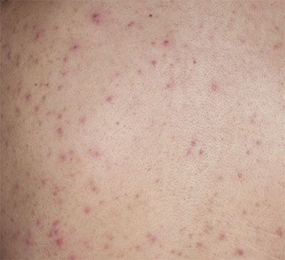
Intensely itchy, hyperpigmented macules developed on the shoulders and upper arms of a 16-year-old boy 2 weeks after he completed his eighth cycle of chemotherapy with bleomycin, etoposide, and cisplatin, following an orchiectomy for a stage IV germ cell tumor of the left testis. During the next 3 days, the lesions evolved into a papulopustular rash that spread to the upper chest, abdomen, and neck.
Intensely itchy, hyperpigmented macules developed on the shoulders and upper arms of a 16-year-old boy 2 weeks after he completed his eighth cycle of chemotherapy with bleomycin, etoposide, and cisplatin, following an orchiectomy for a stage IV germ cell tumor of the left testis. During the next 3 days, the lesions evolved into a papulopustular rash that spread to the upper chest, abdomen, and neck.
A complete blood cell count revealed an absolute neutrophil count of 100/μL. Gram stain of the lesions was negative for bacteria. After a dermatology consultation, Pityrosporum folliculitis was diagnosed on the basis of the clinical findings.
Pityrosporum folliculitis is caused by the fungus Malassezia furfur. Although Malassezia is known to produce a number of chemical compounds that cause relative hypopigmentation of the skin affected by the yeast, hyperpigmented lesions are also frequently seen, as in this case.

Classically described as an acneiform eruption, Malassezia folliculitis appears as 1- to 2-mm, erythematous, monomorphic, pruritic papules and pustules on the upper trunk, neck, and upper arms, and less frequently on the face.1,2 Predisposing factors are causes of immunosuppression, such as HIV infection, organ transplant, and chemotherapy; diabetes mellitus; and use of broad-spectrum antibiotics.1 The differential diagnosis includes bacterial folliculitis, Candida folliculitis (as a manifestation of disseminated candidal infection), acne vulgaris, eosinophilic folliculitis, and graft versus host disease in transplant recipients.1,3 Malassezia folliculitis should be considered in adolescents with acne that fails to respond to traditional therapies.2
Malassezia folliculitis is diagnosed on the basis of clinical picture, microscopy, and response to therapy. Microscopic examination of pustules with a potassium hydroxide preparation shows budding yeast forms and spores, rather than the hyphal or filamentous forms seen in Candida folliculitis.1,3
Treatment usually involves topical antifungals; however, these agents may not penetrate deep enough into the follicles to act on the organism, and oral agents may be required. If oral antifungals are given for more than 6 weeks, liver enzyme levels must be monitored. Recurrence is common; maintenance antifungal regimens (oral ketoconazole once weekly or ketoconazole 2% shampoo 2 or 3 times weekly) may be used as prevention.1 This patient was given a topical 2% ketoconazole cream for 3 weeks. At follow-up, the lesions had begun to resolve.
References:
REFERENCES: 1. Levin NA. Beyond spaghetti and meatballs: skin diseases associated with the Malassezia yeasts. Dermatol Nurs. 2009;21:7-13, 51.
2. Ayers K, Sweeney SM, Wiss K. Pityrosporum folliculitis: diagnosis and management in 6 female adolescents with acne vulgaris. Arch Pediatr Adolesc Med. 2005;159:64-67.
3. Morrison VA, Weisdorf DJ. The spectrum of Malassezia infections in the bone marrow transplant population. Bone Marrow Transplant. 2000;26:645-648.
Recognize & Refer: Hemangiomas in pediatrics
July 17th 2019Contemporary Pediatrics sits down exclusively with Sheila Fallon Friedlander, MD, a professor dermatology and pediatrics, to discuss the one key condition for which she believes community pediatricians should be especially aware-hemangiomas.