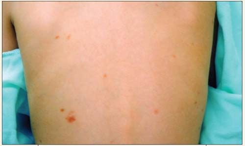
What Rash Consists of These Brownish Macules?
A 4-year-old boy who is new to your practice presents for a well-child visit. His parents report that he has had brownish patches on his torso and back since early infancy. The lesions have decreased in size and number as he has aged. The rash is intermittently pruritic, especially when anyone touches the individual lesions.

THE CASE: A 4-year-old boy who is new to your practice presents for a well-child visit. His parents report that he has had brownish patches on his torso and back since early infancy. The lesions have decreased in size and number as he has aged. The rash is intermittently pruritic, especially when anyone touches the individual lesions. The child has no systemic complaints. The parents deny any vomiting, failure to thrive, developmental concerns, or wheezing.
Answer and Discussion on Next Page
Answer: Urticaria pigmentosa.

Discussion: Urticaria pigmentosa is the most common form of cutaneous mastocytosis. The rash consists of round, red to brown macules or papules that range in number from a few to several thousands. The rash can occur anywhere on the body. The typical lesions of urticaria pigmentosa commonly present within the first 2 years of life.1 When the lesions are stroked, they can become pruritic and erythematous, which is referred to as the Darier sign. The cause of this reaction is mast cell degranulation in response to physical stimulation. Some patients experience more severe systemic symptoms of mast cell degranulation, including flushing, dyspnea, wheezing, nausea, vomiting, and syncope. The natural history of urticaria pigmentosa in children is spontaneous resolution, usually at puberty; however, adolescents with urticaria pigmentosa are less likely to outgrow the symptoms and more likely to have systemic involvement.2
The diagnosis is typically made by the history and physical findings alone, although a skin biopsy can be useful in atypical cases. Treatment is supportive, with oral antihistamines and topical corticosteroids.3
Atopic dermatitis is a chronic relapsing skin disorder characterized by pruritus, xerosis, and papules and vesicles overlying erythematous skin. The rash of histiocytosis (a proliferative disorder of bone marrow derived from Langerhan cells) appears as an eruption of plaques and erythematous papules that may involve petechiae. Epidermolysis bullosa is an inherited disorder that presents in infancy as blister formation at the site of mechanical trauma. The parents confirmed that the child’s rash was not bullous at any time.
References:
REFERENCES:
1. Enta T. Dermacase. Urticaria pigmentosa. Can Fam Physician. 1995;41:1163-1164.
2. Islas AA, Penaranda E. Generalized brownish macules in infancy. Urticaria pigmentosa. Am Fam Physician. 2009;80:987.
3. Correia O, Duarte AF, Quirino P, et al. Cutaneous mastocytosis: two pediatric cases treated with topical pimecrolimus. Dermatol Online J. 2010;16:8.
Recognize & Refer: Hemangiomas in pediatrics
July 17th 2019Contemporary Pediatrics sits down exclusively with Sheila Fallon Friedlander, MD, a professor dermatology and pediatrics, to discuss the one key condition for which she believes community pediatricians should be especially aware-hemangiomas.
Lebrikizumab improves atopic dermatitis symptoms in patients previously on dupilumab
Published: March 7th 2025 | Updated: March 7th 2025Lebrikizumab improves itch, sleep interference, and skin pain in atopic dermatitis patients previously treated with dupilumab, offering a new treatment option.