A 7-Year-Old Boy With Blistering Skin
This 7-year-old boy was recently brought to my office having received a diagnosis of pemphigus foliaceus. His parents were seeking a second opinion.

Case 1:
This 7-year-old boy was recently brought to my office having received a diagnosis of pemphigus foliaceus. His parents were seeking a second opinion.
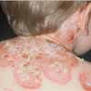
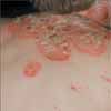
The child had been experiencing superficial blistering of his skin over the past 3 to 4 months: scaling and crusting had developed over the blisters. The eruption involved the patient's upper trunk and face. Small blisters were scattered over his trunk and arms.
The diagnostic test was not a skin biopsy. What was it?

Case 1: This child suffered from extensive bullous impetigo. The confirmatory test was a bacterial culture, which revealed the presence of Staphylococcus aureus.
S aureus produces an exfoliative toxin that results in a cleavage plane in the most superficial layers of the epidermis. This leads to very superficial blisters that easily denude to reveal a glistening surface. If the lesions are untreated, scales and crusts begin to pile up and the surface remains eroded and oozing. Once treatment has eliminated the staphylococci, the skin will rapidly reepithelialize without scarring. Treatment is directed at the staphylococcal organism.
Pemphigus foliaceus is an autoimmune primary blistering disease. The clinical presentation can be very similar to that of bullous impetigo: the epidermal cleavage plane is at an identical level in the epidermis. Both conditions result from the targeting of an epidermal cell adhesion molecule--desmoglein 1--which is responsible for cell-to-cell adhesion in the superficial epidermis. The skin biopsy is often not helpful in differentiating these conditions (the histopathology may be identical).
Direct immunofluorescence testing of the biopsy specimen is diagnostic of pemphigus foliaceus if it shows immunoglobulins surrounding the keratinocytes in the superficial layers of the epidermis. Treatment is directed at the immunologic process with corticosteroids--either topical or systemic--and/or immunosuppressants.

Case 2:
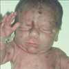
This neonate was born with multiple dull blue-red to brown-red papules and nodules that are accentuated about the head and neck. There are also a number of ecchymotic patches and petechiae over the trunk.
These rare cutaneous findings are classic for what disease?

Case 2: This presentation is classic for blueberry muffin baby. This photograph should be committed to memory.
The term "blueberry muffin baby" is most commonly applied to those circumstances in which there is extramedullary (dermal) erythropoiesis as a result of an intrauterine infection (toxoplasmosis, rubella, Cytomegalovirus infection, or syphilis). Classically, the associated clinical findings and maternal history are significant factors in the diagnosis.
In my opinion, the skin biopsy is the essential diagnostic tool in this setting, and it must be performed during the initial assessment. The differential diagnosis includes congenital leukemia: approximately 50% of neonates with acute myeloid leukemia present with congenital skin lesions. Neuroblastoma and rhabdomyosarcoma are also diagnostic possibilities.
This unfortunate child had congenital myeloid leukemia. The disease was suggested at the "bedside" using a touch preparation of the biopsy specimen before the actual nature of the infiltrate was appreciated. The diagnosis was confirmed by a pediatric oncologist.