- Consultant for Pediatricians Vol 5 No 4
- Volume 5
- Issue 4
Photoclinic: Inflamed Keratosis Pilaris
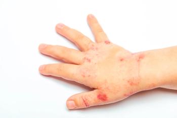
These pinpoint pustules, some with excoriations, and surrounding erythema appeared on the posterior trunk (A) and outer arms (B) of a 15-year-old boy after he had wrapped his upper body in a wool blanket. These lesions were occasionally pruritic, especially on the arms, where most of the excoriations were noted.
Figure A
Figure B
These pinpoint pustules, some with excoriations, and surrounding erythema appeared on the posterior trunk (A) and outer arms (B) of a 15-year-old boy after he had wrapped his upper body in a wool blanket. These lesions were occasionally pruritic, especially on the arms, where most of the excoriations were noted.
Robert P. Blereau, MD, of Morgan City, La, writes that uninflamed keratosis pilaris consists of abundant fine papules and is usually asymptomatic. When inflamed, as in this patient, the papules may become pustular and painful. Aggravating environmental factors include cold, dry air; hot baths; and wool clothing. Scratching, tight-fitting clothes, and abrasive treatments can cause secondary staphylococcal infection.
The diagnosis of keratosis pilaris is based on the clinical appearance of the lesions. This relatively common hereditary disorder typically affects the outer upper arms and anterior and lateral thighs. The condition may be associated with ichthyosis, atopic dermatitis, hyperandrogenism, obesity, and type 1 diabetes mellitus. The differential diagnosis includes acne vulgaris, miliaria, drug-induced eruption, lichen spinulosus, pityriasis rubra pilaris, psoriasis, adverse effects of lithium therapy, and uremia.1
Although there is no cure for keratosis pilaris, the rash may be controlled with topical keratolytics and avoidance of aggravating factors.2
This patient was treated with an oral antistaphylococcal antibiotic (cephalexin) for 10 days and was admonished to avoid contact with abrasive materials. The inflammation promptly resolved.
References:
REFERENCES:
1.
Weedon D.
Skin Pathology.
2nd ed. London: Churchill Livingstone; 2002:482-483.
2.
Blereau RP. Keratosis pilaris.
Consultant.
2004;44:1021.
Articles in this issue
over 19 years ago
Day-Old Boy With Respiratory Distress After Complicated Deliveryover 19 years ago
A 7-Year-Old Boy With Blistering Skinover 19 years ago
Guest Commentary: If I Have Herpes, Can I Still Have Children? . . .over 19 years ago
Neonatal Acne in a 3-Week-Old Boyover 19 years ago
Case in Point: A Young Girl With Cafe au Lait Spotsover 19 years ago
Henoch-Schönlein Purpura and Nonpitting EdemaNewsletter
Access practical, evidence-based guidance to support better care for our youngest patients. Join our email list for the latest clinical updates.


