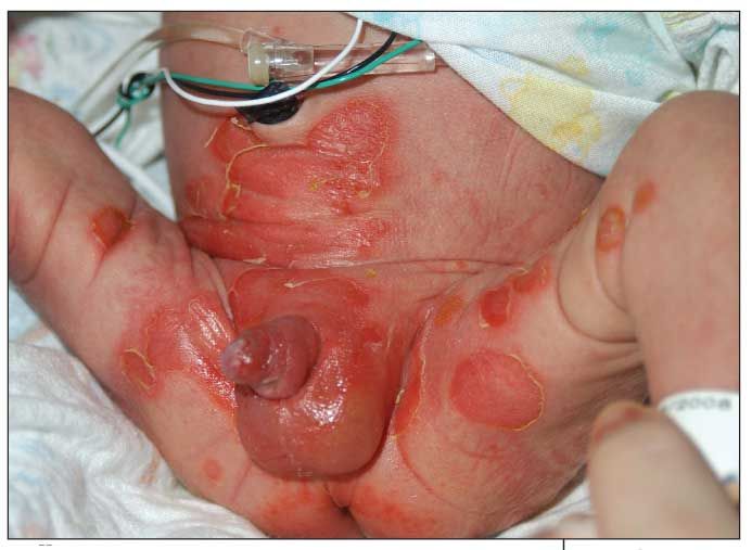
Bullous Impetigo in a 1-Week-Old Boy
Bullous impetigo caused multiple areas of denuded skin, consisting of ruptured bullae with underlying erythema, on the face, lower abdomen, and thighs in this infant.
A 1-week-old boy was brought to an outpatient pediatric clinic by his mother because of concerns of enlarging, peeling lesions around the groin, buttocks, and proximal legs of 2 days’ duration. According to the mother, the lesions appeared to be pus-filled.
The neonate was born at 37 weeks’ gestation via emergent cesarean delivery because of breech presentation. The nursery course was uneventful. The infant remained afebrile; he underwent circumcision and received hepatitis B vaccination before discharge. Prenatal test results were positive for group B streptococcus, but the mother did not receive antibacterial treatment before delivery. There was no family history of bullous disorders.
The infant was admitted to a nearby children’s hospital for further evaluation and management. At that time, the differential diagnosis included toxic epidermal necrolysis, staphylococcal scalded skin syndrome, herpes simplex virus infection, candidiasis, transient neonatal pustulosis, bullous impetigo, and epidermolysis bullosa.

On examination, the infant appeared well-nourished and nontoxic. Vital signs were stable. Multiple areas of denuded skin, consisting of ruptured bullae with underlying erythema, were noted on the face, lower abdomen, and inner thighs-in addition to the groin, buttocks, and proximal legs. One yellow intact bulla of about 1 cm was noted on the left inner thigh. Yellow eye discharge was noted bilaterally; the sclerae and conjunctiva were otherwise normal. All other physical findings were unremarkable.
The patient was treated with ampicillin and cefotaxime. On hospital day 2, dermatology was consulted. Culture of fluid swabbed from an intact blister revealed Staphylococcus aureus. Of the 8 antibiotics for which the organism was tested, it was resistant only to penicillin G. Dermatological diagnosis was bullous impetigo based on clinical criteria and culture results. The patient was treated with cefazolin while in the hospital. He was discharged after 5 days. Outpatient treatment included oral cephalexin and topical mupirocin ointment.
This infant’s rash was typical of bullous impetigo. Whether exposure to the causative organism occurred in the nursery (during or after circumcision) or at home could not be determined. Other possible diagnoses were ruled out on the basis of the following criteria: inconsistency of the rash with both herpes simplex infection and candidiasis and lack of exposure to either infection; lack of toxin/drug exposure; and lack of Nikolsky sign and fever.
Systemic therapy is used in some patients with disseminated lesions. Effective antibiotics include dicloxacillin, amoxicillin plus clavulanic acid, clindamycin, azithromycin, clarithromycin, and cephalosporin. Topical mupirocin is applied 3 times daily for 7 to 10 days, in addion to systemic therapy. Because of the emergence of methicillin-resistant S aureus, clindamycin and trimethoprim/sulfamethoxazole may be used as outpatient therapy.1
Complications of bullous impetigo are rare but may include cellulitis, lymphangitis, suppurative lymphadenitis, guttate psoriasis, and scarlet fever (following streptococcal disease).1 This infant’s rash resolved uneventfully.
Prompt identification of the cause of the rash, whether by clinical presentation, culture and Gram stain of lesion via biopsy, or antibiotic sensitivity testing, is important for appropriate management.
References:
REFERENCE:1. Long SS, Pickering LK, Prober CG, eds. Principles and Practice of Pediatric Infectious Diseases. 3rd ed. New York: Churchill Livingstone; 2008:435-436, 444-445.
Recognize & Refer: Hemangiomas in pediatrics
July 17th 2019Contemporary Pediatrics sits down exclusively with Sheila Fallon Friedlander, MD, a professor dermatology and pediatrics, to discuss the one key condition for which she believes community pediatricians should be especially aware-hemangiomas.