Subclavian Central Venous Catheter Fracture and Embolization
The imaging studies shown are from 2 children with cancer who underwent placement of 9.6 French left subclavian central venous catheters (CVCs) to facilitate treatment. Fracture of the catheters with subsequent embolization of the distal fragment to the pulmonary arteries was noted at about 18 months after placement. Findings suggestive of impending fracture were missed in previous radiographs. In both cases, an interventional radiologist removed the fragment via percutaneous catheterization of the right femoral vein.
The imaging studies shown are from 2 children with cancer who underwent placement of 9.6 French left subclavian central venous catheters (CVCs) to facilitate treatment. Fracture of the catheters with subsequent embolization of the distal fragment to the pulmonary arteries was noted at about 18 months after placement. Findings suggestive of impending fracture were missed in previous radiographs. In both cases, an interventional radiologist removed the fragment via percutaneous catheterization of the right femoral vein.
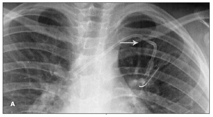
Figure A is from an 8-year-old boy with acute lymphoblastic leukemia. Fluoroscopy confirmed the catheter tip to be at the junction of the superior vena cava and right atrium with no kinks. At about month 19, during an attempt to access the CVC, the patient had pain in his left shoulder. A chest radiograph revealed a fractured piece of the CVC tubing overlying the left lung in the aortopulmonary window region. The distal fragment was lodged in the left main pulmonary artery. The patient could not recall, nor did he complain of, any traumatic events or symptoms that could be attributed to the fracture. On review of previous chest films, an intermittent focal narrowing of the catheter tubing-“pinch-off sign” (POS)-was found. The catheter fracture site (Figure A, arrow) corresponded with the area where the POS was noted.

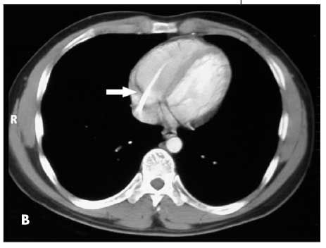
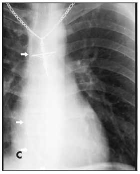
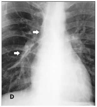
Figures B, C, and D are from a 17-year-old boy with Ewing sarcoma of the left hemipelvis. Fluoroscopy was used to confirm catheter placement, and blood return and ease of flushing the catheter were verified. During a routine clinic visit 18 months later, the patient complained of a cough and “feeling funny.” A chest radiograph was read as normal. A CT scan of the chest, obtained for routine evaluation of possible metastasis, was also interpreted as normal. However, embedded in the radiologist’s report was the comment, “A unipolar transvenous pacemaker has been placed with its tip in the right ventricular apex.” What the radiologist noticed was actually a fractured piece of CVC tubing (Figure B, arrow). Careful inspection of the patient’s earlier “normal” chest radiograph showed the fractured piece of tubing within the right atrium and ventricle (Figure C, arrow). A second chest radiograph showed the tubing fragment lodged in the right pulmonary artery extending into the right lung (Figure D, arrows). No POS was seen in a review of the patient’s radiographic records.


Catheter fracture and embolization of the fractured distal tubing is a rare, lesser-known risk of long-term central venous catheterization. Major risks associated with long-term CVC use are infection, obstruction, and deep venous thrombosis.1 Fracture with embolization should be watched for in any patient with an indwelling CVC. This complication is known to occur in children of all ages, including infants.2 When a long-term CVC is inserted percutaneously into the subclavian vein, the incidence of fracture with distal embolization is 1.6%, with a range of 0.1% to 2.1%.3 This commonly occurs between 6 months and 1 year after implantation.4 At our institution, we had 2 such occurrences in 132 CVCs implanted between 2000 and 2008, for an incidence of 1.5%.
Fracture with embolization may present with signs or symptoms as serious as cardiac arrhythmia5 or as an incidental finding on routine chest imaging. The POS may be the earliest radiographic indication of impending catheter fracture. The POS is thought to occur in 1% of long-term subclavian catheters, with an associated risk of fracture estimated at 40%.6 A significant contributing factor is the gradual breakdown of the tubing secondary to compression between the clavicle and first rib as the tubing passes toward the subclavian vein through the tissue in the costoclavicular space.7
Common sites for embolization of the distal fragment are the central veins, right atrium, right ventricle, and pulmonary arteries. Surprisingly, most patients are asymptomatic. The most common symptom is pain or pressure during an attempt to use the damaged catheter.8 Other common symptoms are cough, palpitations, and swelling around the port site caused by fluid extravasation. Sustained ventricular tachycardia, although rare, may also occur.5 Retrieval of the distal fragment is commonly via a percutaneous vascular route; in some cases, open thoracotomy or even electing to leave the fragment in place may be preferable.9
In the first case, a POS was present in several of the patient’s chest films but was missing in others. This is probably because of the positioning of his arm. When evaluating patients with a CVC and possible intermittent POS, radiographs taken with the arms in adduction and abduction may need to be compared.10 Placing the arms in these positions at the time of insertion may also reveal sites of impingement under fluoroscopy.3 Awareness of the POS allows for replacement of the catheter, if deemed necessary, and spares the patient the risk of complications.
The second case highlights the necessity of careful inspection of all radiographic images ordered and their respective reports. The “funny feeling” the patient had been experiencing may have been related to cardiac arrhythmias caused by stimulation of the right ventricle by the detached tubing fragment.
References:
REFERENCES
:
1.
Kapadia S, Parakh R, Grover T, Yadav A. Catheter fracture and cardiac migration of a totally implantable venous device.
Indian J Cancer.
2005;42:155-157.
2.
Courcoux MF, Jouvet P, Bonnet D, et al. Intravascular rupture of a central venous catheter in a premature infant: retrieval by a nonsurgical technique [in French].
Arch Pediatr.
2000;7:267-270.
3.
Mirza B, Vanek VW, Kepensky DT. Pinch-off syndrome: case report and collective review of the literature.
Am Surg.
2004;70:635-644.
4.
Biffi R, Orsi F, Grasso F, et al. Catheter rupture and distal embolisation: a rare complication of central venous ports.
J Vasc Access.
2000;1:19-22.
5.
Gowda MR, Gowda RM, Khan IA, et al. Positional ventricular tachycardia from a fractured mediport catheter with right ventricular migration-a case report.
Angiology.
2004;55:557-560.
6.
Schlangen JT, Debets JM, Wils JA. The “pinch-off phenomenon”: a radiological symptom for potential fracture of an implanted permanent subclavian catheter system.
Eur J Radiol.
1995;20:112-113.
7.
Hinke DH, Zandt-Stastny DA, Goodman LR, et al. Pinch-off syndrome: a complication of implantable subclavian venous access devices.
Radiology.
1990;177:353-356.
8.
Cheng CC, Tsai TN, Yang CC, Han CL. Percutaneous retrieval of dislodged totally implantable central venous access system in 92 cases: experience in a single hospital.
Eur J Radiol.
2009;69:346-350.
9.
Hayari L, Yalonetsky S, Lorber A. Treatment strategy in the fracture of an implanted central venous catheter.
J Pediatr Hematol Oncol.
2006;28:160-162.
10.
Adwan H, Gordon H, Nicholls E. Are routine chest radiographs needed after fluoroscopically guided percutaneous insertion of central venous catheters in children?
J Pediatr Surg.
2008;43:341-343.
Newsletter
Access practical, evidence-based guidance to support better care for our youngest patients. Join our email list for the latest clinical updates.