A Collage of Genital Lesions, Part 4
A discussion of three types of Genital Lesions: Sebaceous Gland Hyperplasia of the Penis, Varicocele, and Penile Dog Bite.

Sebaceous Gland Hyperplasia of the Penis
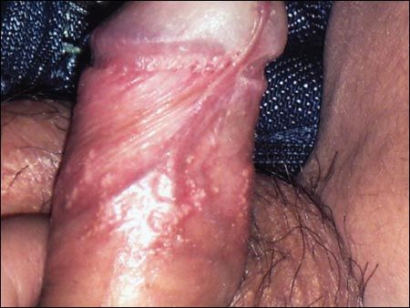
These papular lesions on the penis of a 14-year-old boy first appeared 1 year before presentation. The lesions had remained unchanged in size and number and were asymptomatic. Urination and penile erection were undisturbed.
On examination, multiple yellowish papules were noted on the dorsal surface of the distal penile shaft. A clinical diagnosis of sebaceous gland hyperplasia of the penis was made.
Also known as sebaceous gland prominence on the penis and ectopic sebaceous glands (Fordyce disease), sebaceous gland hyperplasia of the penis typically presents as soft, yellowish papules, 1 to 3 mm in diameter, on the penile shaft. The condition is asymptomatic.
Histologically, the sebaceous glands in the upper dermis in the penile shaft are hyperplastic, with lobules of varying sizes. The clinical differential diagnosis includes sebaceous adenoma, milia, lichen planus, lichen nitidus, pearly penile papules, and warts.
The patient was reassured of the benign nature of the condition and that no treatment was required. The lesions were still present after 2 years.
FOR MORE INFORMATION:
■ Carson HJ, Massa M, Reddy V. Sebaceous gland hyperplasia of the penis.
J Urol
. 1996;156:1441.
■ Kumar A, Kossard S. Band-like sebaceous hyperplasia over the penis.
Australas J Dermatol
. 1999;40:47-48.
■ Vergara G, Belinchón I, Silvestre JF, et al. Linear sebaceous gland hyperplasia of the penis: a case report.
J Am Acad Dermatol
. 2003;48:149-150.

Varicocele
A 12-year-old boy noticed this mass in the left side of the scrotum a few months earlier. The mass enlarged when the child was in an upright position and when he performed the Valsalva maneuver. It was asymptomatic. Urination was normal. There was no recent weight loss. Dilated veins were visible on the overlying skin.
On examination, the mass felt like a bag of worms, which is characteristic of a varicocele.
A varicocele results from incompetent valves in the spermatic vein. Incompetent valves permit transmission of hydrostatic venous pressure to the pampiniform plexus. The resulting distention and tortuosity of the pampiniform plexus are so obvious that a varicocele is unlikely to be confused with any other condition.
Varicoceles are rare in children younger than 10 years, except in those with Wilms tumor, neuroblastoma, or hydronephrosis that causes obstruction of venous return from the testis. In contrast, the incidence in adolescent boys has been reported to be 5% to 15%. The left side is affected in about 90% of cases, presumably because the left pampiniform plexus and spermatic vein drain into the left renal vein, whereas the right spermatic vein drains into a lower point in the inferior vena cava. In 10% of cases, the condition is bilateral. Varicoceles may present with slight discoloration (as in this case), as a result of the distention and tortuosity of the pampiniform plexus.
The abnormality is usually asymptomatic and is often detected during a routine physical examination. However, a large varicocele may cause a dragging sensation or dull, aching pain, particularly during strenuous exercise. As in this boy, a varicocele is typically most prominent when the patient is in a standing position and enlarges with the Valsalva maneuver. Because of the increase in scrotal temperature, a varicocele may lead to a low sperm count or reduced sperm motility and may interfere with fertility. Some affected boys may have a reduction in testosterone production or impaired gonadotropin response to gonadotropin-releasing hormone stimulation.
Once a varicocele is present, it tends to persist throughout the boy's lifetime. If a varicocele is a suspected cause of infertility, it may be treated by surgical ligation of the spermatic vein (eg, with laparoscopic spermatic vein clipping). Although the effect of early surgical correction of a varicocele on future fertility is unknown, improved testicular growth has been reported after surgical correction in adolescents. Other indications for surgery are pain, testicular growth retardation or arrest over 6 to 12 months, a varicocele in a solitary testis, and a marked volume disparity between the testicles.
FOR MORE INFORMATION:
■ Leung AKC, Wong AL. Pediatric scrotal swelling.
Consultant for Pediatricians
. 2003;2:172-176.
■ MacLellan DL, Diamond DA. Recent advances in external genitalia.
Pediatr Clin North Am
. 2006;53:449-464, vii.

Penile Dog Bite
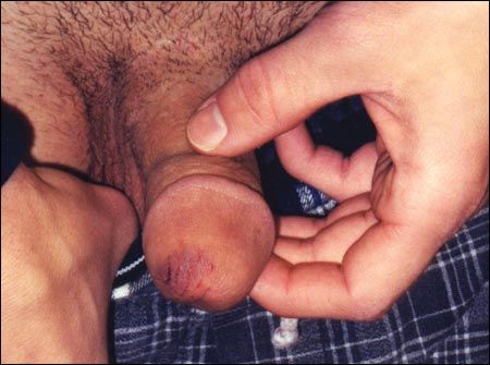
A 16-year-old boy presented with wounds on the penis after he was bitten by the family dog in the bathroom. Urination was unaffected. The boy's immunization status was up-to-date for tetanus. The dog was fully immunized for rabies.
The patient had 2 tiny puncture marks on the glans penis with some bruising. There were no signs of infection. His wounds were cleaned with copious amounts of sterile saline, and he recovered uneventfully. No prophylactic antibiotic was prescribed.
Penile dog bites may lead to localized bacterial infection, penile disfiguration, voiding dysfunction, or sexual dysfunction. About 5% to 10% of dog bite injuries become infected. The most common causative organisms are Pasteurella multocida and Staphylococcus aureus. Most infections manifest as cellulitis or an abscess.
The recommended treatment includes thorough wound cleansing and careful debridement of necrotic or devitalized tissue. Primary wound closure is preferred when no obvious infection is present. Otherwise, the wound can be approximated and allowed to close by delayed primary or secondary intention.
Routine use of a prophylactic antibiotic without obvious signs of infection is controversial. When choosing between an oral and parenteral antibiotic for an established wound infection, consider the severity of the wound, the degree of overt infection, signs of toxicity, and the patient's immune status. Amoxicillin/clavulanate is the drug of choice for empiric oral therapy. Ticarcillin/clavulanate or ampicillin/sulbactam is preferred for patients who require empiric parenteral therapy.
FOR MORE INFORMATION:
■ Gomes CM, Ribeiro-Filho L, Giron AM, et al. Genital trauma due to animal bites.
J Urol
. 2000;165:80-83.
■ Leung AK, Robson WL. Penile dog bite in an adolescent.
Adv Ther
. 2005;22:363-367.
Recognize & Refer: Hemangiomas in pediatrics
July 17th 2019Contemporary Pediatrics sits down exclusively with Sheila Fallon Friedlander, MD, a professor dermatology and pediatrics, to discuss the one key condition for which she believes community pediatricians should be especially aware-hemangiomas.