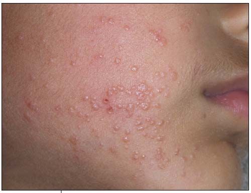
Molluscum Contagiosum on the Face of a 4-Year-Old Boy
Despite curettage several months earlier, the facial rash on this 4-year-old boy had spread across both cheeks and was now mildly pruritic.

Despite curettage several months earlier, the facial rash on this 4-year-old boy had spread across both cheeks and was now mildly pruritic. These discrete, smooth, pearly or flesh-colored, dome-shaped papules with central umbilication are characteristic of molluscum contagiosum.
This benign viral infection, first described in 1817, is caused by a double-stranded DNA virus in the Poxviridae family. It affects healthy children between 2 and 11 years old, although those with immunodeficiency, atopic disease, and Darier disease are at increased risk for the condition.1,2 This patient’s growth charts showed that he was thriving. Transmission is through close physical contact, autoinoculation, and fomites (such as clothing and towels).3 The incubation period is 2 to 7 weeks.4
The infection occurs in the superficial epidermis, occasionally of the mucous membranes, and conjunctiva.5 The lesions typically measure 1 to 5 mm in diameter and number fewer than 30.1 However, they can be larger and extensive in immunocompromised patients. The lesions are most often located on the trunk, axillae, and antecubital and popliteal fossae. Children with molluscum contagiosum involving the genitals should be examined for possible sexual abuse. The rash is usually asymptomatic but can itch or become irritated if picked or scratched. Secondary bacterial infection is a rare complication.
Diagnosis is made on the distinctive clinical appearance. The differential diagnoses include common warts, papular acrodermatitis of childhood, papular urticaria, and varicella.
Although most lesions resolve spontaneously, some patients request active treatment, as opposed to watching and waiting, for social and aesthetic reasons or because of concerns about spreading the disease to others, as was the case for this patient.6 Evidence from comparative trials suggests no single best treatment method.7 Active treatment may be mechanical (curettage, cryotherapy with liquid nitrogen), chemical (tretinoin, cantharidin, podophyllotoxin), or immunological (imiquimod, cimetidine).
This patient was treated with imiquimod 1% cream. The cream was applied gently on individual papules every other night for 2 weeks. The rash resolved without scarring.
References:
REFERENCES:
1. Gottlieb SL, Myskowski PL. Molluscum contagiosum. Int J Dermatol. 1994; 33:453-461.
2. Brown J, Janniger CK, Schwartz RA, Silverberg NB. Childhood molluscum contagiosum. Int J Dermatol. 2006;45:93-99.
3. Diven DG. An overview of poxviruses. J Am Acad Dermatol. 2001;44:1-16.
4. Tyring SK. Molluscum contagiosum: the importance of early diagnosis and treatment. Am J Obstet Gynecol. 2003;189(3 suppl):S12-S16
5. Silverberg NB. Warts and molluscum in children. Adv Dermatol. 2004;20: 23-73.
6. Jones S, Kress D. Treatment of molluscum contagiosum and herpes simplex virus cutaneous infections. Cutis. 2007;79(4 suppl):11-17.
7. van der Wouden JC, Menke J, Gajadin S, et al. Interventions for cutaneous molluscum contagiosum. Cochrane Database Syst Rev. 2006;(2):CD0004767.
Recognize & Refer: Hemangiomas in pediatrics
July 17th 2019Contemporary Pediatrics sits down exclusively with Sheila Fallon Friedlander, MD, a professor dermatology and pediatrics, to discuss the one key condition for which she believes community pediatricians should be especially aware-hemangiomas.