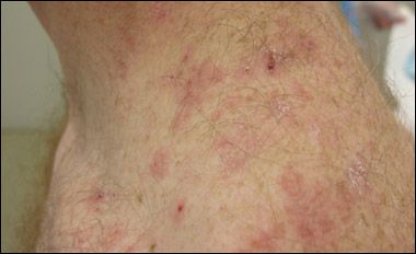
Atypical Dermatitis Herpetiformis
An 18-year-old boy presented with a several-month history of an intermittent, very pruritic rash on his back that did not improve with topical corticosteroids. Physical examination revealed grouped erythematous papules with a few scattered small vesicles on his posterior neck and bilateral posterior shoulders at the location where his backpack frequently rubbed.

An 18-year-old boy presented with a severalmonth history of an intermittent, very pruritic rash on his back that did not improve with topical corticosteroids. Physical examination revealed grouped erythematous papules with a few scattered small vesicles on his posterior neck and bilateral posterior shoulders at the location where his backpack frequently rubbed. A biopsy of the lesion with immunofluorescence studies showed subepidermal separation with collections of neutrophils at the dermal papillary tips and granular IgA deposits in the dermal papillae. On the basis of the biopsy results, dermatitis herpetiformis was diagnosed.
Dermatitis herpetiformis is an uncommon skin condition that usually presents in young adults but can develop at any time. It is a classic cutaneous manifestation of glutensensitive enteropathy, or celiac disease-also referred to as "celiac sprue." The rash is often intermittent, usually coinciding with dietary exposure to gluten.
The typical presentation involves grouped vesicles and papules concentrated on the extensor elbows, knees, and buttocks, often with overlying excoriations secondary to the marked pruritus. The rash tends to occur in areas of pressure; in this case, the diagnosis was difficult because the shoulders are not usually considered areas of pressure and it is unusual for dermatitis herpetiformis to present there. In children, dermatitis herpetiformis may have an atypical appearance and resemble atopic dermatitis, scabies, impetigo, or papular urticaria.1 This case illustrates the variable clinical presentations of dermatitis herpetiformis, especially in younger patients.
Only 20% of persons with dermatitis herpetiformis have GI complaints, although 90% have evidence of intestinal involvement on further evaluation.2
Patients with celiac disease may also complain of arthritis, peripheral neuropathy, ataxia, anemia, and recurrent aphthous ulcers. In addition, they have an increased risk of other autoimmune disorders, including thyroid disease, autoimmune hepatitis, type 1 diabetes, and psoriasis.3 Perhaps the most serious sequela is the increased risk of intestinal and extraintestinal lymphomas.
The diagnosis of dermatitis herpetiformis can be established in several ways. Clinical suspicion is important, but a skin biopsy with direct immunofluorescence of uninvolved skin is the gold standard for confirmation of the diagnosis. A less invasive study-testing for IgA antiepidermal transglutaminase antibodies-detects serological evidence of dermatitis herpetiformis and is very sensitive. Testing for anti-tissue transglutaminase antibodies can reveal a correlation with the degree of intestinal involvement and can be used to document adherence to a gluten-free diet.4 However, intestinal involvement is confirmed by a small-bowel biopsy.
Treatment of the rash includes the use of potent topical corticosteroids, which are usually only partially effective. Oral dapsone is very effective and can relieve itching in a few days; however, it can also lead to relapse on discontinuation.1 Dapsone should not be used in patients with glucose-6-phosphate dehydrogenase deficiency because of the risk of hemolytic anemia. Although dapsone is very effective at clearing the rash, treatment should center on adherence to a gluten-free diet because the risk of subsequent lymphoma development is not decreased by dapsone therapy.3
On further questioning, this patient did report frequent episodes of diarrhea. Both the rash and GI symptoms resolved over several months with adherence to a gluten-free diet.
References:
REFERENCE:
1.
Caproni M, Antiga E., Melani L, Fabbri P; Italian Group for Cutaneous Immunopathology. Guidelines for the diagnosis and treatment of dermatitis herpetiformis.
J Eur Acad Dermatol Venereol
. 2009;23:633-638.
2.
Bolognia JL, Jorizzo JL, Rapini RP, eds.
Dermatology
. Elsevier; 2008:447-452.
3.
Hernandez L, Green PH. Extraintestinal manifestations of celiac disease.
Curr Gastroenterol Rep
. 2006;8:383-389.
4.
Rose C, Armbruster FP, Ruppert J, et al. Autoantibodies against epidermal transglutaminase are a sensitive diagnostic marker in patients with dermatitis herpetiformis on a normal or gluten-free diet.
J Am Acad Dermatol
. 2009;61:39-43.
Recognize & Refer: Hemangiomas in pediatrics
July 17th 2019Contemporary Pediatrics sits down exclusively with Sheila Fallon Friedlander, MD, a professor dermatology and pediatrics, to discuss the one key condition for which she believes community pediatricians should be especially aware-hemangiomas.