Toddler With Nonpruritic Rash That Does Not Respond to Corticosteroids
A 1-year-old boy presented with a 10-day history of a nonpruritic rash that had persisted and spread despite treatment with a topical corticosteroid. Mother reported that he was febrile at the onset of the eruption; he was given over-the-counter antipyretics. On day 3, his pediatrician evaluated his condition and prescribed amoxicillin for his fever and hydrocortisone cream for his atopic dermatitis. Over the next several days, the fever subsided; however, the rash, which had started on the child’s right hand, persisted and spread to his face and elsewhere.

HISTORY
A 1-year-old boy presented with a 10-day history of a nonpruritic rash that had persisted and spread despite treatment with a topical corticosteroid. Mother reported that he was febrile at the onset of the eruption; he was given over-the-counter antipyretics. On day 3, his pediatrician evaluated his condition and prescribed amoxicillin for his fever and hydrocortisone cream for his atopic dermatitis. Over the next several days, the fever subsided; however, the rash, which had started on the child's right hand, persisted and spread to his face and elsewhere.
The child had been relatively healthy. He had a history of atopic dermatitis, but he had no allergies and had had no notable illnesses.
PHYSICAL EXAMINATION
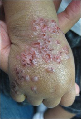
Patient was afebrile and generally well-appearing. Examination of the skin revealed punched-out erosions and crusted erythematous papules on dorsum of right hand and wrist, and erythematous papules on right side of face, right anterior shoulder, and left dorsal wrist. The skin elsewhere on his body had a normal appearance. All other physical findings were normal.
"WHAT'S YOUR DIAGNOSIS?"
Answer on Next Page
ANSWER: ECZEMA HERPETICUM

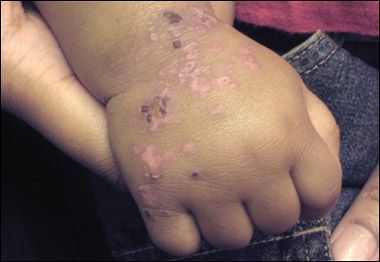
Figure – Here, the child's hand is seen after 1 week of treatment with oral acyclovir. Most of the vesicles have resolved.
Atopic dermatitis is one of the most common chronic skin conditions seen by both primary care pediatricians and pediatric dermatologists. Eczema herpeticum (EH) is a cutaneous superinfection with herpes simplex virus (HSV) that develops in patients with atopic dermatitis; it requires immediate attention. EH is the most common form of Kaposi varicelliform eruption, a broader term that refers to secondary viral infections that develop in preexisting skin disorders (such as eczema, pemphigus, ichthyosis, burns); the causative organisms include HSV type 1, vaccinia virus, and coxsackieviruses.1,2 In atopic dermatitis, the xerosis and intense pruritus result in a disrupted skin barrier with a predisposition to secondary infection by numerous microorganisms, including staphylococcus, molluscum, and HSV.
CLINICAL MANIFESTATIONS
EH is seen more frequently in infants and small children but may present at any age. Initially, clusters of multiple, uniform, 2- to 3-mm crusted papules and vesicles develop in untreated or poorly controlled atopic dermatitis. The eruption progresses rapidly, and the vesicles rupture and form punched-out ulcerations. The patient often experiences increased pain and burning at the site of the eruption.
EH may occur on any part of the body but is more common on the upper extremities and face and is confined to areas of eczematous skin.2 The initial infection may be associated with a prodrome of fever, malaise, and lymphadenopathy.
DIFFERENTIAL DIAGNOSIS
EH is frequently confused with impetigo, scabies, varicella, herpes zoster, and an exacerbation of the underlying atopic dermatitis. Unlike EH, varicella, herpes zoster, and scabies are not simply confined to areas of underlying dermatitis. Also, the punched-out ulcerations of EH are characteristic of the disorder and can help distinguish it from atopic dermatitis flares and impetigo.
DIAGNOSIS
The diagnosis of EH is clinical. Confirmatory tests are available. However, if testing is pursued, antiviral therapy should be initiated at the time of clinical suspicion and not be delayed for pending results. Of the tests available, viral culture has the highest sensitivity and specificity; however, culture results can take several hours to days to be considered final. Results of direct fluorescent antibody testing may be available more quickly, but clinicians who perform this test need to be knowledgeable about the proper specimen collection technique. An adequate specimen-one that provides sufficient epithelial cells for diagnosis-must be obtained by scraping the base of an unroofed vesicle.
TREATMENT
Therapy with acyclovir should be started promptly in any patient in whom EH is suspected. Topical or oral antibiotics for bacterial superinfection, most commonly staphylococcal, should be initiated as indicated.
Patients with mild, localized symptoms may be treated on an outpatient basis with oral acyclovir. However, close clinical monitoring to ensure improvement is of paramount importance.
Any child with lesions in close proximity to the eye or with visual complaints should be examined by an ophthalmologist to rule out herpetic keratitis, which is considered an ophthalmological emergency and requires immediate therapy to prevent progression.
Children who are febrile or ill-appearing, as well as young infants, should be hospitalized and given parenteral acyclovir, intravenous hydration, and pain management. Also, consider a workup for systemic sepsis in such patients; laboratory studies recommended for evaluation of systemic infection include complete blood cell count, blood urea nitrogen level, creatinine level, and aminotransferase levels.3
Because this boy was afebrile and well-appearing, he was given oral acyclovir; after 1 week, this therapy had produced significant resolution of the rash (Figure).
DISEASE COURSE
Lesions usually persist for several weeks.4 The incidence of acyclovir-resistant HSV is extremely low; nonetheless, patients should be re-evaluated in several days to ensure adequate response. Although repair of the epidermal barrier is the ultimate goal, which in patients with atopic dermatitis might normally require treatment with corticosteroids and aggressive use of emollients, therapy with oral or topical corticosteroids may theoretically cause progression of the HSV infection. Thus, use of these agents should be postponed until antiviral therapy has been completed and all active vesiculopustular lesions have resolved. In the interim, oral antihistamines and emollients that do not contain corticosteroids can be used.
Children are considered infectious until all lesions have crusted over. Those enrolled in school should keep any active lesions covered to reduce the risk of transmission.5
Although symptoms tend to be milder in recurrences of EH, children with recurring HSV infection may benefit from preventive therapy in the form of lower-dose daily oral acyclovir, at least until their atopic dermatitis has been brought under tighter clinical control.
COMPLICATIONS
If treatment of EH is delayed, severe complications may develop; these can include systemic HSV infection, sepsis from secondary bacterial infection, and permanent visual deficits as a result of ocular keratitis. Thus, it is important to have a high index of clinical suspicion to detect and promptly treat EH.
References:
REFERENCE:
1.
Vandana Madkan V, Karan S, Brantley J, et al. Human herpesviruses. In: Bolognia JL, Jorizzo JL, Rapini RP, eds.
Dermatology
. 2nd ed. London: Elsevier; 2008.
2.
Olson J, Robles DT, Kirby P, Colven R. Kaposi varicelliform eruption (eczema herpeticum).
Dermatol Online J
. 2008;14:18.
3.
Mackley CL, Adams DR, Anderson B, Miller JJ. Eczema herpeticum: a dermatologic emergency.
Dermatol Nurs
. 2002;14:307-310, 323; quiz, 313.
4.
Viral infections of skin and mucosa. In: Wolff K, Johnson RA, Suurmond D.
Fitzpatrick's Color Atlas and Synopsis of Clinical Dermatology
. 5th ed. New York: McGraw-Hill Companies; 2005.
5.
American Academy of Pediatrics. Herpes simplex. In: Pickering LK, Baker CJ, Long SS, McMillan JA, eds.
Red Book: 2006 Report of the Committee on Infectious Diseases
. 27th ed. Elk Grove, IL: American Academy of Pediatrics; 2006.
Recognize & Refer: Hemangiomas in pediatrics
July 17th 2019Contemporary Pediatrics sits down exclusively with Sheila Fallon Friedlander, MD, a professor dermatology and pediatrics, to discuss the one key condition for which she believes community pediatricians should be especially aware-hemangiomas.